Shi Xiangguang, Xia Xueyi, Xiao Yang, Zhang Ying, Gong Yiyi, Chen Yahui, Shi Chenyi, Wang Wei, Liu Jianlan, Huang Jia, Liu Mengguo, Xu Zhuoya, Ma Yanyun, Shi Mengkun, Wang Jiucun, Wu Wenyu
Department of Dermatology, Huashan Hospital, Fudan University, Shanghai, China.
Department of Dermatology, State Key Laboratory of Molecular Engineering of Polymers, Shanghai Institute of Dermatology, Huashan Hospital, Jing'an District Central Hospital, Fudan University, Shanghai, China.
Cell Commun Signal. 2025 Mar 18;23(1):141. doi: 10.1186/s12964-025-02116-z.
Keloid is a typical skin fibrotic disease with unclear mechanisms and limited therapeutic options. Fibroblast-induced fibrogenesis is a crucial cause of KD. However, the types of cells involved in fibroblast fibrogenesis in KD and the specific mechanisms are unclear. This study aimed to investigate the role of melanocyte-secreted melanin in promoting fibroblast fibrogenesis and its mechanism and to evaluate the potential therapeutic effect of intervening melanin in treating keloid.
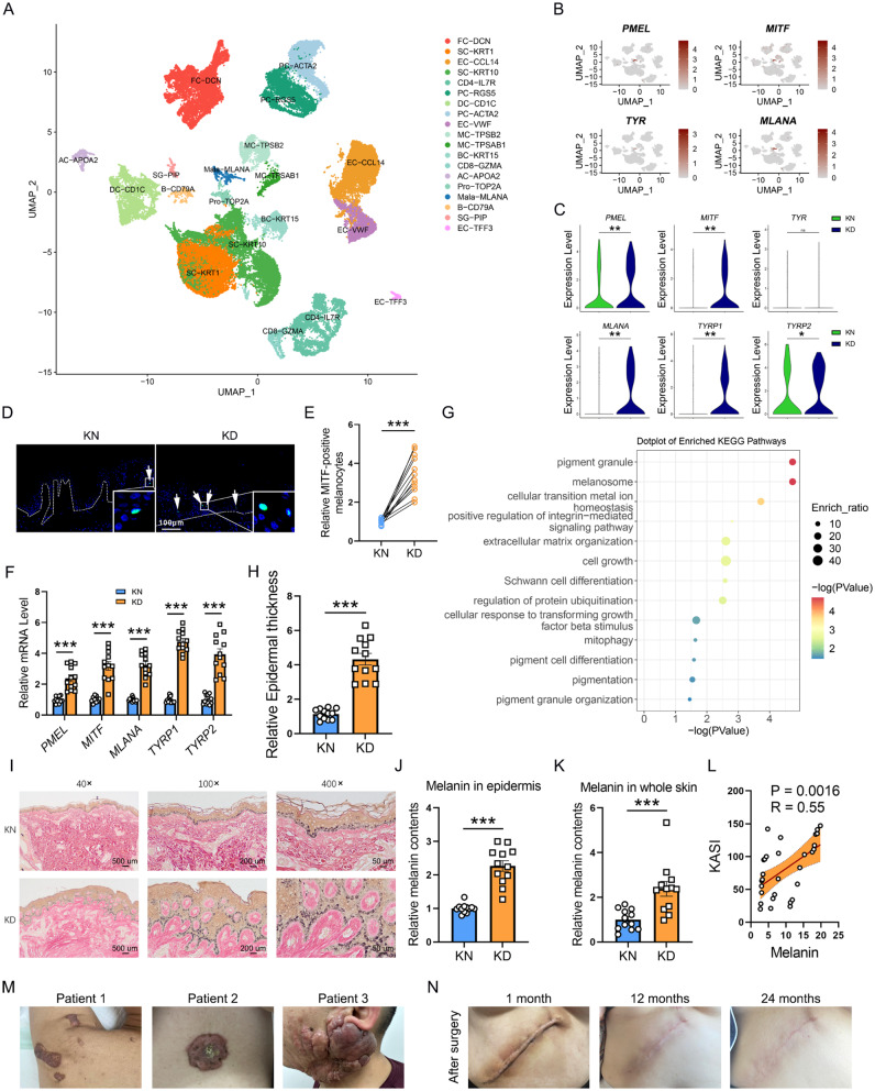
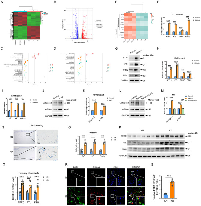
The activity of pigmentation-related pathways in KD melanocytes was examined using single-cell RNA-sequence (scRNA-seq) analysis. Masson-Fontana staining or isolated melanin quantification detected the melanin levels and distribution in the skin and cells. Collagen deposition, wounding healing, and proliferation analysis were employed to integratively assess fibroblast fibrogenesis. After melanin treatment, bulk-seq identified fibroblasts' differentially expressed genes (DEGs). The iron levels were detected by Perl's staining or isolated iron quantification. Cell viability, LipidROS, and malondialdehyde assay accessed the ferroptosis levels. The therapeutic potential of ML329 was evaluated in keloid-bearing mice.
We found the enriched skin pigmentation-related pathways in the melanocytes of keloid by single-cell RNA-sequence (scRNA-seq) analysis. We further validated increased melanin levels in keloid patients. Additionally, melanin positively correlated with the Keloid Area and Severity Index in keloid. Furthermore, melanocyte-secreted melanin significantly promoted fibroblast proliferation, migration, and collagen synthesis. Mechanically, melanin increased basal cell permeability and inflammation to facilitate its transfer to the dermis, where it further activated fibroblasts by evoking iron overload and ferroptosis resistance. Consistently, iron overload and ferroptosis resistance were validated in primary fibroblasts and skin tissues of keloid patients. Inhibition of iron overload and ferroptosis resistance effectively diminish melanin-induced fibrogenesis. Interestingly, melanin induced iron overload and ferroptosis resistance in melanocytes in an autocrine manner and further stimulated keratinocytes to take up melanin to deepen skin color by upregulating the F2R-like trypsin receptor 1 (F2RL1). In vivo, the delivery of ML329, a microphthalmia-associated transcription factor (MITF) inhibitor, could suppress melanogenesis and alleviate keloid in human keloid-bearing nude mice. Meanwhile, ML329 decreased the iron content and restored the sensitivities of ferroptosis.
Collectively, melanin-lowing strategies may appear as a potential new therapeutic target for keloid.
瘢痕疙瘩是一种典型的皮肤纤维化疾病,其发病机制尚不清楚,治疗选择有限。成纤维细胞诱导的纤维化是瘢痕疙瘩的一个关键病因。然而,瘢痕疙瘩中成纤维细胞纤维化过程中涉及的细胞类型及具体机制尚不清楚。本研究旨在探讨黑素细胞分泌的黑色素在促进成纤维细胞纤维化中的作用及其机制,并评估干预黑色素在治疗瘢痕疙瘩中的潜在治疗效果。
使用单细胞RNA测序(scRNA-seq)分析检测瘢痕疙瘩黑素细胞中色素沉着相关信号通路的活性。采用Masson-Fontana染色或分离的黑色素定量检测皮肤和细胞中的黑色素水平及分布。采用胶原沉积、伤口愈合及增殖分析综合评估成纤维细胞纤维化。黑色素处理后,批量测序鉴定成纤维细胞的差异表达基因(DEG)。通过Perl染色或分离的铁定量检测铁水平。通过细胞活力、脂质活性氧和丙二醛测定评估铁死亡水平。在瘢痕疙瘩小鼠中评估ML329的治疗潜力。
通过单细胞RNA测序(scRNA-seq)分析,我们发现瘢痕疙瘩黑素细胞中存在丰富的皮肤色素沉着相关信号通路。我们进一步验证了瘢痕疙瘩患者黑色素水平升高。此外,黑色素与瘢痕疙瘩的瘢痕疙瘩面积和严重程度指数呈正相关。此外,黑素细胞分泌的黑色素显著促进成纤维细胞增殖、迁移和胶原合成。机制上,黑色素增加基底细胞通透性和炎症,以促进其转移至真皮,在真皮中它通过引发铁过载和铁死亡抗性进一步激活成纤维细胞。一致地,在瘢痕疙瘩患者的原代成纤维细胞和皮肤组织中验证了铁过载和铁死亡抗性。抑制铁过载和铁死亡抗性可有效减少黑色素诱导的纤维化。有趣的是,黑色素以自分泌方式诱导黑素细胞中的铁过载和铁死亡抗性,并通过上调F2R样胰蛋白酶受体1(F2RL1)进一步刺激角质形成细胞摄取黑色素以加深肤色。在体内,递送小眼畸形相关转录因子(MITF)抑制剂ML329可抑制黑色素生成并减轻人瘢痕疙瘩裸鼠的瘢痕疙瘩。同时,ML329降低铁含量并恢复铁死亡敏感性。
总体而言,降低黑色素的策略可能成为瘢痕疙瘩潜在的新治疗靶点。