Xu Can, Nie Xinyu, Xu Ru, Zhou Luyang, Wang Dongjin
Department of Cardiac Surgery, Nanjing Drum Tower Hospital, Nanjing University Medical School, Nanjing, People's Republic of China.
Nanjing University Medical School, Nanjing, People's Republic of China.
Tob Induc Dis. 2025 Mar 18;23. doi: 10.18332/tid/201400. eCollection 2025.
We aimed to explore the role of Apelin-13 in resisting oxidation, inflammation as well as apoptosis and its underlying mechanisms of action using a model of nicotine-induced H9c2 cardiomyocyte injury.
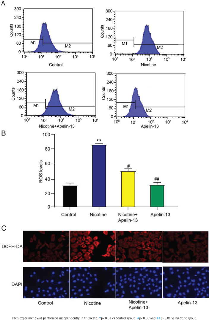
H9c2 cardiomyocytes were randomly divided into control, nicotine, nicotine + Apelin-13, and Apelin-13 groups. Cell counting kit-8 assay was conducted to determine the cell viability. Interleukin (IL)-6, superoxide dismutase, tumor necrosis factor-alpha (TNF-α), glutathione peroxidase (GSH-Px), IL-β, catalase (CAT), IL-8, lactate dehydrogenase (LDH), and malondialdehyde (MDA) levels were examined. A 2',7'-dichlorodihydrofluorescein diacetate assay was conducted to measure the intracellular reactive oxygen species (ROS) level. The morphology of apoptotic cardiomyocytes was observed by 4',6-diamidino-2-phenylindole staining. Western blotting was employed to measure the protein expressions of apoptotic factors B-cell lymphoma-2 (Bcl-2) and Bcl-2-associated X (Bax). Apoptosis was quantified using Annexin V/propidium iodide staining.
Exposure of H9c2 cardiomyocytes to 10 μM nicotine significantly reduced cell viability and increased LDH release, oxidative stress (elevated MDA and ROS levels with decreased superoxide dismutase, GSH-Px, and CAT activities), pro-inflammatory cytokines (IL-6, TNF-α, IL-1β, IL-8), and apoptotic markers (increased Bax with decreased Bcl-2 expression, along with nuclear condensation) (p<0.05). In contrast, treatment with 2 μM Apelin-13 significantly alleviated these deleterious effects, enhancing cell viability, restoring antioxidant enzyme activities, reducing oxidative and inflammatory responses, and inhibiting apoptosis (p<0.05).
Nicotine induction increases the oxidative stress and apoptotic capacity of H9c2 cardiomyocytes, but Apelin-13 protects H9c2 cardiomyocytes against nicotine-induced apoptosis and oxidative stress.
我们旨在利用尼古丁诱导的H9c2心肌细胞损伤模型,探讨Apelin-13在抗氧化、抗炎以及抗凋亡方面的作用及其潜在作用机制。
将H9c2心肌细胞随机分为对照组、尼古丁组、尼古丁+Apelin-13组和Apelin-13组。采用细胞计数试剂盒-8法检测细胞活力。检测白细胞介素(IL)-6、超氧化物歧化酶、肿瘤坏死因子-α(TNF-α)、谷胱甘肽过氧化物酶(GSH-Px)、IL-1β、过氧化氢酶(CAT)、IL-8、乳酸脱氢酶(LDH)和丙二醛(MDA)水平。采用2',7'-二氯二氢荧光素二乙酸酯法检测细胞内活性氧(ROS)水平。通过4',6-二脒基-2-苯基吲哚染色观察凋亡心肌细胞的形态。采用蛋白质印迹法检测凋亡因子B细胞淋巴瘤-2(Bcl-2)和Bcl-2相关X蛋白(Bax)的蛋白表达。使用膜联蛋白V/碘化丙啶染色对凋亡进行定量分析。
将H9c2心肌细胞暴露于10μM尼古丁中可显著降低细胞活力,并增加LDH释放、氧化应激(MDA和ROS水平升高,同时超氧化物歧化酶、GSH-Px和CAT活性降低)、促炎细胞因子(IL-6、TNF-α、IL-1β、IL-8)以及凋亡标志物(Bax增加,Bcl-2表达降低,同时伴有核浓缩)(p<0.05)。相比之下,用2μM Apelin-13处理可显著减轻这些有害影响,提高细胞活力,恢复抗氧化酶活性,减少氧化和炎症反应,并抑制凋亡(p<0.05)。
尼古丁诱导可增加H9c2心肌细胞的氧化应激和凋亡能力,但Apelin-13可保护H9c2心肌细胞免受尼古丁诱导的凋亡和氧化应激。