Liu Yushuang, Duan Zhengyu, Luo Zhongzhou, Zhang Runze, Li Jiaxiong, Zhang Jinze, Meng Zeyu, Wang Bowen, Yuan Jin, Xiao Peng
State Key Laboratory of Ophthalmology, Zhongshan Ophthalmic Centre, Guangdong Provincial Key Laboratory of Ophthalmology and Visual Science, Guangdong Provincial Clinical Research Centre for Ocular Diseases, Sun Yat-Sen University, Guangzhou, China.
Ophthalmol Sci. 2025 Jan 12;5(3):100712. doi: 10.1016/j.xops.2025.100712. eCollection 2025 May-Jun.
Conjunctival goblet cells (CGCs) play a crucial role in maintaining ocular surface health by producing mucins. However, assessing CGC changes in ocular diseases remains limited by invasive techniques and subjective evaluations. This study aims to develop a noncontact cellular resolution fluorescence microscopy for in vivo CGC imaging and investigate CGC dynamics in a dry eye disease (DED) mouse model.
Experimental study.
Freshly ex vivo porcine eyes, New Zealand white rabbits, and C57BL/6 mice.
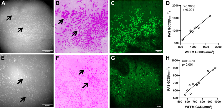
Based on the intrinsic fluorescence properties of moxifloxacin, a high-resolution noncontact widefield fluorescence microscopy (WFFM) was customized with an all-in-focus algorithm to optimize in vivo CGC imaging over the curved conjunctival surface. A DED mouse model was established by topically applying 0.2% benzalkonium chloride (BAC) to the ocular surface daily for 7 days, followed by a 7-day recovery period without BAC. In vivo CGC alterations were assessed using WFFM on days 0, 3, 7, and 14. Additional assessments included the phenol red thread tear test, corneal sodium fluorescein staining, and periodic acid-Schiff (PAS) assay.
Conjunctival goblet cell density and area ratio.
The WFFM system achieved a cellular resolution of 1 μm and a field of view of 1.4 mm × 1.4 mm. Imaging validation in mice and rabbits allowed for the distinguishing and quantitative assessment of individual CGCs or clusters on the curved conjunctival surface in vivo. Significant reductions in CGC density and area ratio on days 3 and 7 after BAC induction were observed in DED mouse in vivo with WFFM, with their values returning to the baseline 7 days after BAC removal, which was consistent with PAS staining results.
The customized WFFM enables in vivo cellular imaging of CGCs, offering a safe and accurate method for continuous monitoring of CGC pathophysiology in ocular surface diseases such as DED.
Proprietary or commercial disclosure may be found in the Footnotes and Disclosures at the end of this article.
结膜杯状细胞(CGCs)通过产生粘蛋白在维持眼表健康中起关键作用。然而,评估眼部疾病中CGCs的变化仍然受到侵入性技术和主观评估的限制。本研究旨在开发一种用于体内CGC成像的非接触式细胞分辨率荧光显微镜,并研究干眼疾病(DED)小鼠模型中的CGC动态变化。
实验研究。
新鲜离体猪眼、新西兰白兔和C57BL/6小鼠。
基于莫西沙星的固有荧光特性,定制了一种高分辨率非接触式宽场荧光显微镜(WFFM),并采用全聚焦算法优化在弯曲结膜表面的体内CGC成像。通过每天在眼表局部应用0.2%苯扎氯铵(BAC)7天建立DED小鼠模型,随后在无BAC的情况下恢复7天。在第0、3、7和14天使用WFFM评估体内CGC的变化。额外的评估包括酚红棉线泪液试验、角膜荧光素钠染色和过碘酸-希夫(PAS)染色。
结膜杯状细胞密度和面积比。
WFFM系统实现了1μm的细胞分辨率和1.4mm×1.4mm的视野。在小鼠和兔子中的成像验证允许在体内对弯曲结膜表面的单个CGC或细胞簇进行区分和定量评估。在DED小鼠体内,用WFFM观察到BAC诱导后第3天和第7天CGC密度和面积比显著降低,在去除BAC 7天后其值恢复到基线水平,这与PAS染色结果一致。
定制的WFFM能够对CGCs进行体内细胞成像,为连续监测DED等眼表疾病中的CGC病理生理学提供了一种安全、准确的方法。
在本文末尾的脚注和披露中可能会找到专有或商业披露信息。