• 文献检索
  • 文档翻译
  • 深度研究
  • 学术资讯
  • Suppr Zotero 插件Zotero 插件
  • 邀请有礼
  • 套餐&价格
  • 历史记录
应用&插件
Suppr Zotero 插件Zotero 插件浏览器插件Mac 客户端Windows 客户端微信小程序
定价
高级版会员购买积分包购买API积分包
服务
文献检索文档翻译深度研究API 文档MCP 服务
关于我们
关于 Suppr公司介绍联系我们用户协议隐私条款
关注我们

Suppr 超能文献

核心技术专利:CN118964589B侵权必究
粤ICP备2023148730 号-1Suppr @ 2026

文献检索

告别复杂PubMed语法,用中文像聊天一样搜索,搜遍4000万医学文献。AI智能推荐,让科研检索更轻松。

立即免费搜索

文件翻译

保留排版,准确专业,支持PDF/Word/PPT等文件格式,支持 12+语言互译。

免费翻译文档

深度研究

AI帮你快速写综述,25分钟生成高质量综述,智能提取关键信息,辅助科研写作。

立即免费体验

CT引导下肺活检时肺高灌注依赖区穿刺路径磨玻璃影及活检作为肺出血危险因素的回顾性研究

Ground-Glass Opacities in the Access Route and Biopsy in Highly Perfused Dependent Areas of the Lungs as Risk Factors for Pulmonary Hemorrhage During CT-Guided Lung Biopsy: A Retrospective Study.

作者信息

Brönnimann Michael P, Manser Leonie, Christe Andreas, Heverhagen Johannes T, Gebauer Bernhard, Auer Timo A, Schnapauff Dirk, Collettini Federico, Schroeder Christophe, Dorn Patrick, Gassenmaier Tobias, Ebner Lukas, Huber Adrian T

机构信息

Department of Diagnostic, Interventional and Paediatric Radiology, Inselspital, Bern University Hospital, University of Bern, Rosenbühlgasse 27, 3010 Bern, Switzerland.

Department of Radiology, Charité-Universitätsmedizin Augustenburger Platz 1, 13353 Berlin, Germany.

出版信息

Tomography. 2025 Mar 14;11(3):35. doi: 10.3390/tomography11030035.

DOI:10.3390/tomography11030035
PMID:40137575
原文链接:https://pmc.ncbi.nlm.nih.gov/articles/PMC11945665/
Abstract

BACKGROUND/OBJECTIVES: The risk of hemorrhage during CT-guided lung biopsy has not been systematically studied in cases where ground-glass opacities (GGO) are present in the access route or when biopsies are performed in highly perfused, dependent lung areas. While patient positioning has been studied for pneumothorax prevention, its role in minimizing hemorrhage risk remains unexplored. This study aimed to determine whether GGOs in the access route and biopsies in dependent lung areas are risk factors for pulmonary hemorrhage during CT-guided lung biopsy.

METHODS

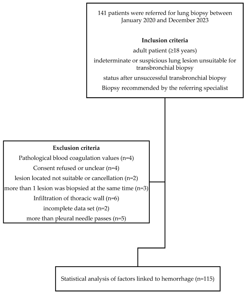
A retrospective analysis was conducted on 115 CT-guided lung biopsies performed at a single center (2020-2023). Patients were categorized based on post-interventional hemorrhage exceeding 2 cm (Grade 2 or higher). We evaluated the presence of GGOs in the access route and biopsy location (dependent vs. non-dependent areas) using chi square, Fisher's exact, and Mann-Whitney U tests. Univariate and multivariate logistic regression analyses were conducted to evaluate risk factors for pulmonary hemorrhage.

RESULTS

Pulmonary hemorrhage beyond 2 cm occurred in 30 of 115 patients (26%). GGOs in the access route were identified in 67% of these cases ( < 0.01), and hemorrhage occurred more frequently when biopsies were performed in dependent lung areas (63% vs. 40%, = 0.03). Multivariable analysis showed that GGOs in the access route (OR 5.169, 95% CI 1.889-14.144, = 0.001) and biopsies in dependent areas (OR 4.064, 95% CI 1.477-11.186, < 0.001) independently increased hemorrhage risk.

CONCLUSIONS

GGOs in the access route and dependent lung area biopsies are independent risk factors for hemorrhage during CT-guided lung biopsy.

摘要

背景/目的:在CT引导下肺活检过程中,当穿刺路径存在磨玻璃影(GGO)或在肺灌注良好的下垂部位进行活检时,出血风险尚未得到系统研究。虽然已经研究了患者体位以预防气胸,但其在降低出血风险方面的作用仍未得到探索。本研究旨在确定穿刺路径中的GGO和下垂肺区活检是否为CT引导下肺活检时肺出血的危险因素。

方法

对在单一中心进行的115例CT引导下肺活检(2020 - 2023年)进行回顾性分析。根据介入后出血超过2 cm(2级或更高)对患者进行分类。我们使用卡方检验、Fisher精确检验和Mann-Whitney U检验评估穿刺路径和活检部位(下垂部位与非下垂部位)是否存在GGO。进行单因素和多因素逻辑回归分析以评估肺出血的危险因素。

结果

115例患者中有30例(26%)发生了超过2 cm的肺出血。这些病例中有67%在穿刺路径中发现了GGO(<0.01),并且在下垂肺区进行活检时出血更频繁(63%对40%,=0.03)。多变量分析显示,穿刺路径中的GGO(OR 5.169,95%CI 1.889 - 14.144,=0.001)和下垂部位活检(OR 4.064,95%CI 1.477 - 11.186,<0.001)独立增加出血风险。

结论

穿刺路径中的GGO和下垂肺区活检是CT引导下肺活检时出血的独立危险因素。

https://cdn.ncbi.nlm.nih.gov/pmc/blobs/71be/11945665/13491c75117a/tomography-11-00035-g008.jpg
https://cdn.ncbi.nlm.nih.gov/pmc/blobs/71be/11945665/f8adcdec2861/tomography-11-00035-g001.jpg
https://cdn.ncbi.nlm.nih.gov/pmc/blobs/71be/11945665/53ab18fae9e7/tomography-11-00035-g002.jpg
https://cdn.ncbi.nlm.nih.gov/pmc/blobs/71be/11945665/aa956e9cdee5/tomography-11-00035-g003.jpg
https://cdn.ncbi.nlm.nih.gov/pmc/blobs/71be/11945665/4d751463eeea/tomography-11-00035-g004.jpg
https://cdn.ncbi.nlm.nih.gov/pmc/blobs/71be/11945665/0444138fb192/tomography-11-00035-g005.jpg
https://cdn.ncbi.nlm.nih.gov/pmc/blobs/71be/11945665/9736e79ceaff/tomography-11-00035-g006.jpg
https://cdn.ncbi.nlm.nih.gov/pmc/blobs/71be/11945665/0e367f8ab690/tomography-11-00035-g007.jpg
https://cdn.ncbi.nlm.nih.gov/pmc/blobs/71be/11945665/13491c75117a/tomography-11-00035-g008.jpg

相似文献

1
Ground-Glass Opacities in the Access Route and Biopsy in Highly Perfused Dependent Areas of the Lungs as Risk Factors for Pulmonary Hemorrhage During CT-Guided Lung Biopsy: A Retrospective Study.CT引导下肺活检时肺高灌注依赖区穿刺路径磨玻璃影及活检作为肺出血危险因素的回顾性研究
Tomography. 2025 Mar 14;11(3):35. doi: 10.3390/tomography11030035.
2
Hemorrhage risk prediction after computed tomography-guided lung biopsy: Clinical parameters and quantitative pulmonary vascular analysis.计算机断层扫描引导下肺活检后的出血风险预测:临床参数与肺血管定量分析
J Formos Med Assoc. 2025 Jan;124(1):79-86. doi: 10.1016/j.jfma.2024.03.011. Epub 2024 Mar 20.
3
Pulmonary Hemorrhage Following Percutaneous Computed Tomography-Guided Lung Biopsy: Retrospective Review of Risk Factors, Including Aspirin Usage.经皮计算机断层扫描引导下肺活检术后肺出血:包括阿司匹林使用情况在内的危险因素回顾性分析
Curr Probl Diagn Radiol. 2020 Jan-Feb;49(1):12-16. doi: 10.1067/j.cpradiol.2018.10.007. Epub 2018 Nov 2.
4
Dependent lesion positioning at CT-guided lung biopsy to reduce risk of pneumothorax.CT 引导下肺活检中依赖病变定位以降低气胸风险。
Eur Radiol. 2020 Nov;30(11):6369-6375. doi: 10.1007/s00330-020-07025-y. Epub 2020 Jun 27.
5
Pneumothorax Complicating Coaxial and Non-coaxial CT-Guided Lung Biopsy: Comparative Analysis of Determining Risk Factors and Management of Pneumothorax in a Retrospective Review of 650 Patients.气胸并发同轴和非同轴CT引导下肺活检:对650例患者进行回顾性分析以确定气胸危险因素及处理的比较研究
Cardiovasc Intervent Radiol. 2016 Feb;39(2):261-70. doi: 10.1007/s00270-015-1167-3. Epub 2015 Jul 7.
6
Pneumothorax risk reduction during CT-guided lung biopsy - Effect of fluid application to the pleura before lung puncture and the gravitational effect of pleural pressure.CT 引导下肺活检中气胸风险的降低——在肺穿刺前向胸膜应用液体和胸膜压力的重力效应的影响。
Eur J Radiol. 2024 Jul;176:111529. doi: 10.1016/j.ejrad.2024.111529. Epub 2024 May 24.
7
Diagnostic accuracy and complications of CT-guided core needle lung biopsy of solid and part-solid lesions.CT 引导下经皮肺穿刺活检术对实性及部分实性病变的诊断准确性及并发症。
Br J Radiol. 2018 Jul;91(1088):20170946. doi: 10.1259/bjr.20170946. Epub 2018 Jun 1.
8
Risk factors for pneumothorax and pulmonary hemorrhage following computed tomography-guided transthoracic core-needle biopsy of subpleural lung lesions.CT 引导下经皮胸膜下肺病变穿刺活检并发气胸和肺出血的危险因素。
J Chin Med Assoc. 2022 Apr 1;85(4):500-506. doi: 10.1097/JCMA.0000000000000705.
9
Risk factor analysis of pulmonary hemorrhage complicating CT-guided lung biopsy in coaxial and non-coaxial core biopsy techniques in 650 patients.650例患者在同轴和非同轴芯针活检技术中CT引导下肺活检并发肺出血的危险因素分析
Eur J Radiol. 2014 Oct;83(10):1945-52. doi: 10.1016/j.ejrad.2014.06.023. Epub 2014 Jul 5.
10
Diagnostic Accuracy and Safety of Nonsurgical Biopsy for Diagnosing Pulmonary Ground-Glass Opacities: A Systematic Review and Meta-Analysis.非手术活检诊断肺部磨玻璃影的诊断准确性和安全性:系统评价和荟萃分析。
Respiration. 2024;103(11):661-674. doi: 10.1159/000539876. Epub 2024 Jul 29.

本文引用的文献

1
Innovative strategies for minimizing hematoma risk in MRI-guided breast biopsies.磁共振成像引导下乳腺活检中降低血肿风险的创新策略。
Radiol Oncol. 2025 Jan 22;59(1):91-99. doi: 10.2478/raon-2025-0004. eCollection 2025 Mar 1.
2
Enhancing safety in CT-guided lung biopsies: correlation of MinIP imaging with pneumothorax risk prediction.提高CT引导下肺活检的安全性:最小强度投影(MinIP)成像与气胸风险预测的相关性
Insights Imaging. 2025 Jan 13;16(1):16. doi: 10.1186/s13244-024-01890-7.
3
Enhanced Positioning Strategies to Reduce Pneumothorax in CT-Guided Lung Biopsies.
减少CT引导下肺活检气胸的强化定位策略
Diagnostics (Basel). 2024 Nov 23;14(23):2639. doi: 10.3390/diagnostics14232639.
4
Incidence and risk factors for pulmonary hemorrhage after percutaneous CT-guided pulmonary nodule biopsy: an observational study.经皮 CT 引导下肺结节活检后肺出血的发生率及危险因素:一项观察性研究。
Sci Rep. 2024 Mar 28;14(1):7348. doi: 10.1038/s41598-024-58045-3.
5
Avoiding the Intercostal Arteries in Percutaneous Thoracic Interventions.避免经皮胸腔介入中的肋间动脉。
J Vasc Interv Radiol. 2022 Apr;33(4):416-419.e2. doi: 10.1016/j.jvir.2021.12.026.
6
The PEARL Approach for CT-guided Lung Biopsy: Assessment of Complication Rate.PEARL 法 CT 引导下肺活检:并发症发生率评估。
Radiology. 2022 Feb;302(2):473-480. doi: 10.1148/radiol.2021210360. Epub 2021 Nov 2.
7
Correlation between ground-glass opacity on pulmonary CT and the levels of inflammatory cytokines in patients with moderate-to-severe COVID-19 pneumonia.肺 CT 磨玻璃密度与中重度 COVID-19 肺炎患者炎症细胞因子水平的相关性。
Int J Med Sci. 2021 Apr 16;18(11):2394-2400. doi: 10.7150/ijms.56683. eCollection 2021.
8
Ground-glass opacity (GGO): a review of the differential diagnosis in the era of COVID-19.磨玻璃密度影(GGO):COVID-19 时代的鉴别诊断综述。
Jpn J Radiol. 2021 Aug;39(8):721-732. doi: 10.1007/s11604-021-01120-w. Epub 2021 Apr 26.
9
Factors influencing early recurrence of hepatocellular carcinoma after curative resection.影响肝癌根治性切除术后早期复发的因素。
J Int Med Res. 2020 Aug;48(8):300060520945552. doi: 10.1177/0300060520945552.
10
Risk factors associated with pulmonary hemorrhage and hemoptysis following percutaneous CT-guided transthoracic lung core needle biopsy: a retrospective study of 1,090 cases.经皮CT引导下经胸肺穿刺活检术后肺出血和咯血的相关危险因素:1090例回顾性研究
Quant Imaging Med Surg. 2020 May;10(5):1008-1020. doi: 10.21037/qims-19-1024.