Cameron Jillian M, Mito Remika, Berkovic Samuel F, Jackson Graeme D
Epilepsy Research Centre, Department of Medicine, University of Melbourne, Austin Health, Melbourne, Victoria, Australia.
Florey Institute of Neuroscience and Mental Health, Heidelberg, Victoria, Australia.
Ann Clin Transl Neurol. 2025 Jun;12(6):1135-1143. doi: 10.1002/acn3.70010. Epub 2025 Apr 10.
Progressive myoclonus epilepsy (PME) is a rare generalized epilepsy syndrome with a well-characterized genetic basis. The brain networks that are affected to give rise to the distinctive symptoms of PME are less well understood.
Eleven individuals with PME with a confirmed genetic diagnosis and 22 controls were studied. MRI included diffusion acquisition using 64 directions, b = 3000 s/mm. Fixel-based analysis was used to identify white matter pathways with significant abnormality in structural connectivity, with subsequent tract segmentation and analysis. Region-of-interest and whole-brain volumetric analysis of T1-weighted images was performed. The relationship between structural connectivity measures and disease duration, and Unified Myoclonus Rating Scale was assessed.
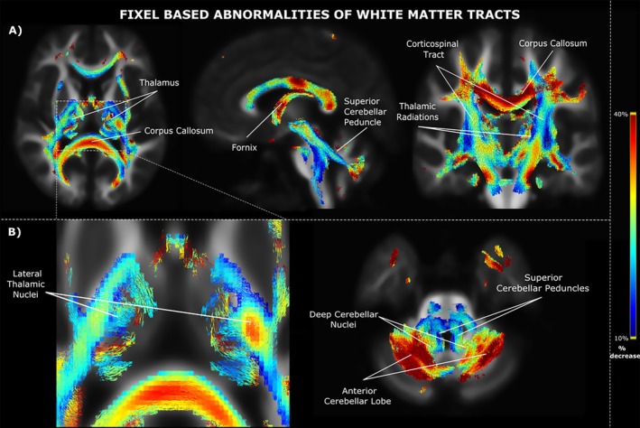
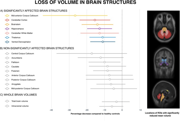
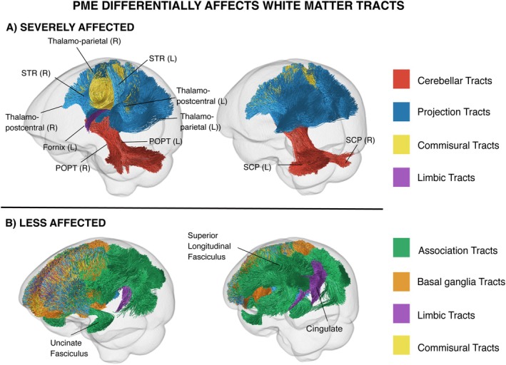
Analysis of structural connectivity revealed significantly reduced fiber density and fiber bundle cross-section in white matter tracts in individuals with PME, with the most severe involvement of tracts within the cerebello-thalamo-cortical network, particularly the cerebello-thalamic, thalamo-cortical, cortico-thalamic, and corticospinal tracts, as well as the splenium of the corpus callosum. By comparison with these abnormalities, cortico-cortical association pathways were relatively preserved. There was reduced volume in the cerebellum, thalamus, brainstem, and mid-anterior corpus callosum.
Individuals with PME have atrophy and changes in fiber tracts that predominantly affect the cerebello-thalamic and motor systems, likely reflecting neuronal and axonal loss as part of a degenerative process. This imaging pattern is distinctive and accords well with the characteristic clinical, neuropathological, and neurophysiological features of the PMEs. The mechanism by which the PME genes affect these tracts is not yet known.
进行性肌阵挛癫痫(PME)是一种罕见的全身性癫痫综合征,具有明确的遗传基础。导致PME独特症状的受影响脑网络尚不太清楚。
对11名经基因确诊的PME患者和22名对照者进行了研究。MRI包括使用64个方向、b = 3000 s/mm²的扩散采集。基于固定点的分析用于识别结构连接性存在显著异常的白质通路,随后进行束分割和分析。对T1加权图像进行感兴趣区域和全脑体积分析。评估结构连接性测量与疾病持续时间以及统一肌阵挛评分量表之间的关系。
结构连接性分析显示,PME患者白质束中的纤维密度和纤维束横截面积显著降低,小脑 - 丘脑 - 皮质网络内的束受累最为严重,特别是小脑 - 丘脑、丘脑 - 皮质、皮质 - 丘脑和皮质脊髓束,以及胼胝体压部。与这些异常相比,皮质 - 皮质联合通路相对保留。小脑、丘脑、脑干和胼胝体中前部体积减小。
PME患者存在萎缩和纤维束变化,主要影响小脑 - 丘脑和运动系统,这可能反映了作为退行性过程一部分的神经元和轴突损失。这种影像学模式是独特的,与PME的特征性临床、神经病理学和神经生理学特征相符。PME基因影响这些束的机制尚不清楚。