• 文献检索
  • 文档翻译
  • 深度研究
  • 学术资讯
  • Suppr Zotero 插件Zotero 插件
  • 邀请有礼
  • 套餐&价格
  • 历史记录
应用&插件
Suppr Zotero 插件Zotero 插件浏览器插件Mac 客户端Windows 客户端微信小程序
定价
高级版会员购买积分包购买API积分包
服务
文献检索文档翻译深度研究API 文档MCP 服务
关于我们
关于 Suppr公司介绍联系我们用户协议隐私条款
关注我们

Suppr 超能文献

核心技术专利:CN118964589B侵权必究
粤ICP备2023148730 号-1Suppr @ 2026

文献检索

告别复杂PubMed语法,用中文像聊天一样搜索,搜遍4000万医学文献。AI智能推荐,让科研检索更轻松。

立即免费搜索

文件翻译

保留排版,准确专业,支持PDF/Word/PPT等文件格式,支持 12+语言互译。

免费翻译文档

深度研究

AI帮你快速写综述,25分钟生成高质量综述,智能提取关键信息,辅助科研写作。

立即免费体验

促炎细胞因子通过反式激活癌细胞上的糖基化细胞因子受体,诱导上皮-间质转化,形成转移表型。

Pro-Inflammatory Cytokines Transactivate Glycosylated Cytokine Receptors on Cancer Cells to Induce Epithelial-Mesenchymal Transition to the Metastatic Phenotype.

作者信息

Baghaie Leili, Bunsick David A, Aucoin Emilyn B, Skapinker Elizabeth, Yaish Abdulrahman M, Li Yunfan, Harless William W, Szewczuk Myron R

机构信息

Department of Biomedical & Molecular Sciences, Queen's University, Kingston, ON K7L 3N6, Canada.

Faculty of Science, Biology (Biomedical Science), York University, Toronto, ON M3J 1P3, Canada.

出版信息

Cancers (Basel). 2025 Apr 5;17(7):1234. doi: 10.3390/cancers17071234.

DOI:10.3390/cancers17071234
PMID:40227834
原文链接:https://pmc.ncbi.nlm.nih.gov/articles/PMC11988151/
Abstract

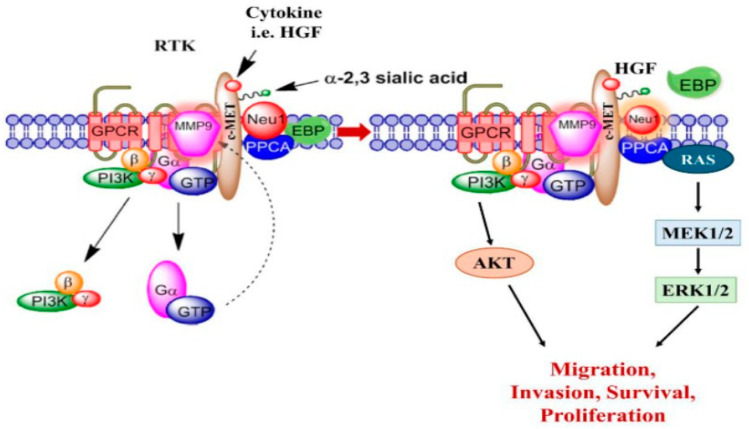
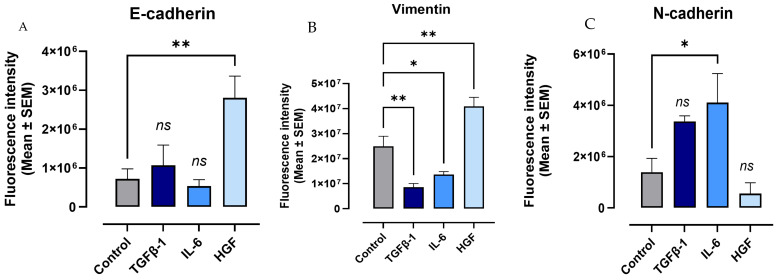
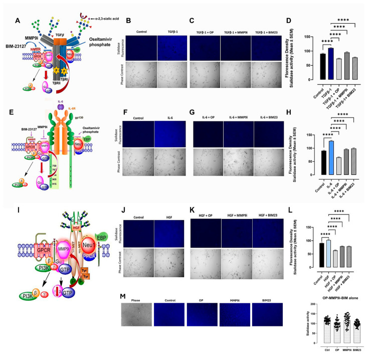
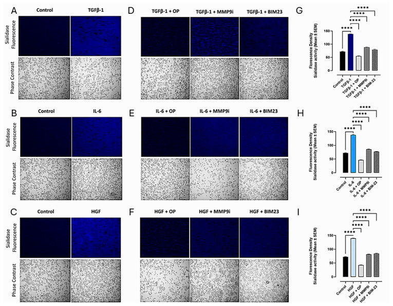
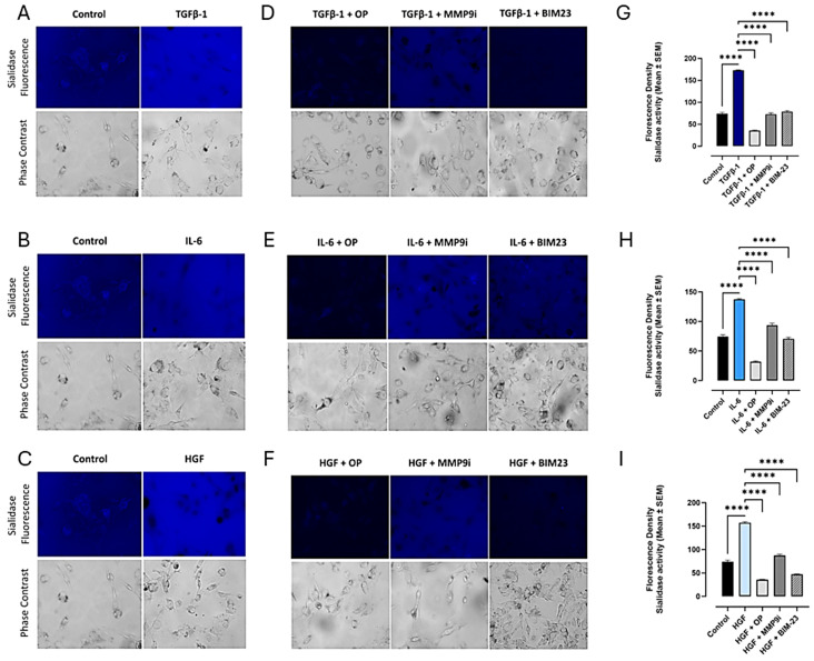
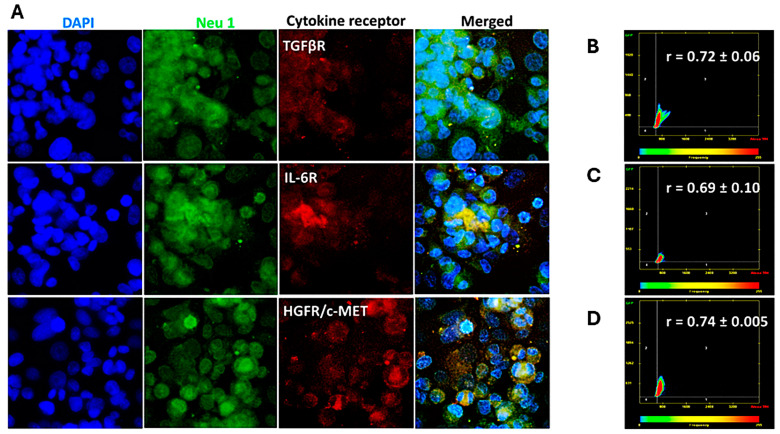
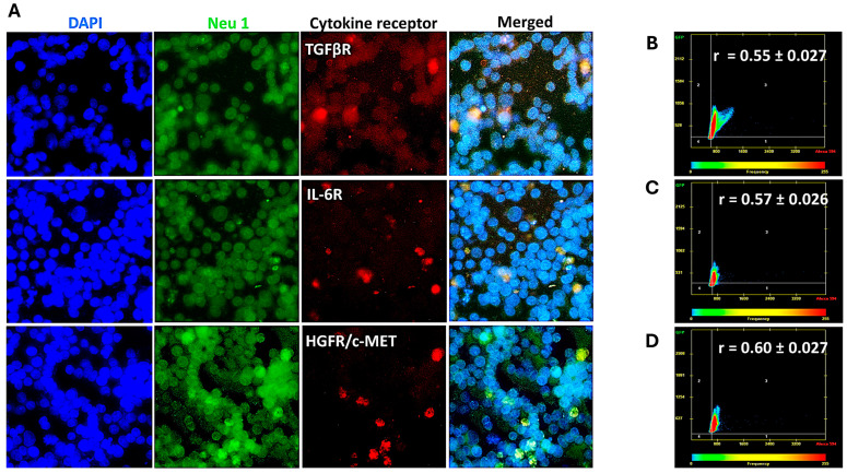
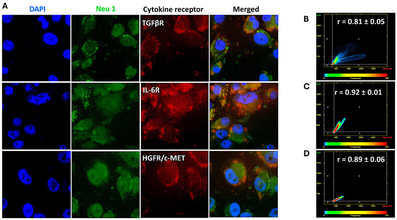
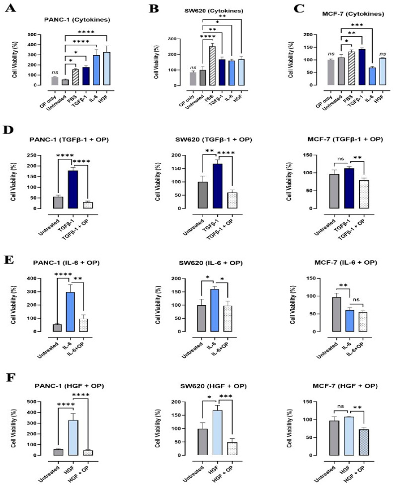
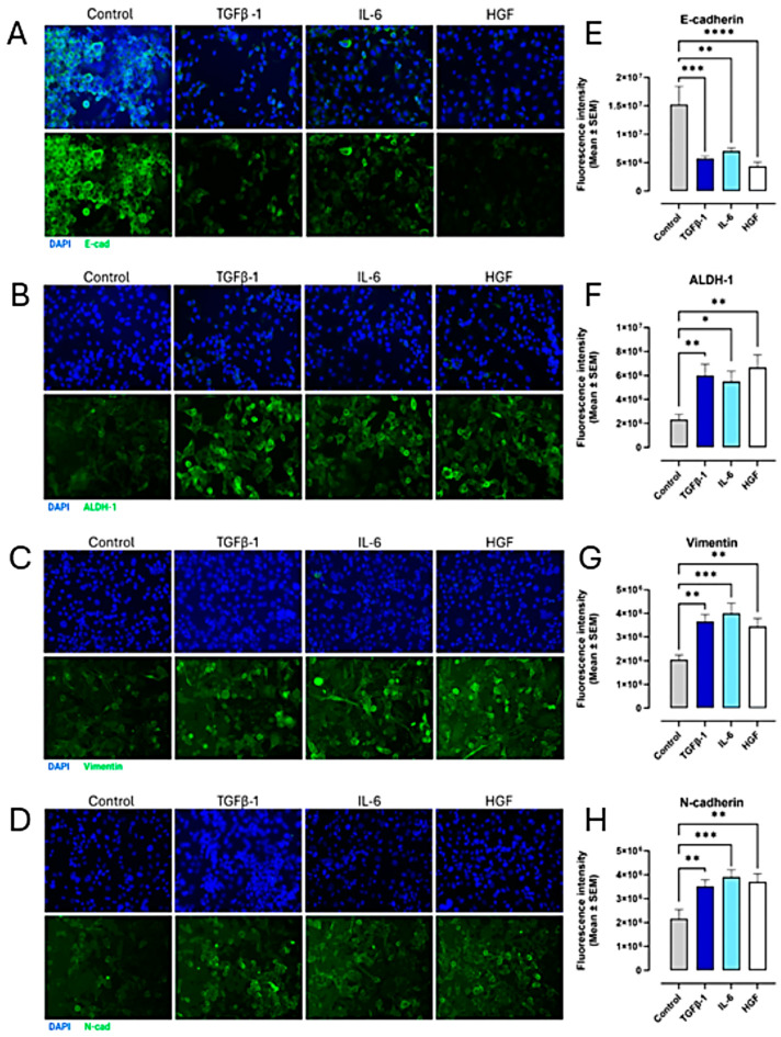
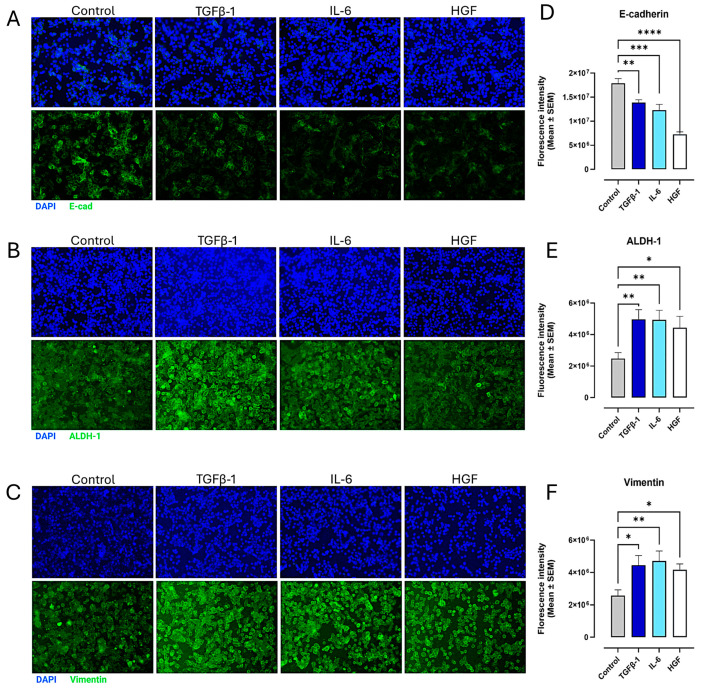
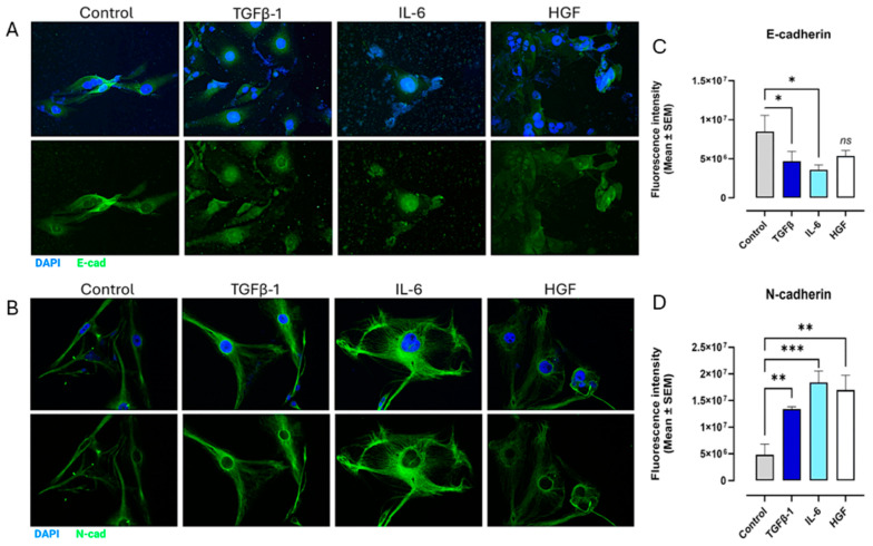
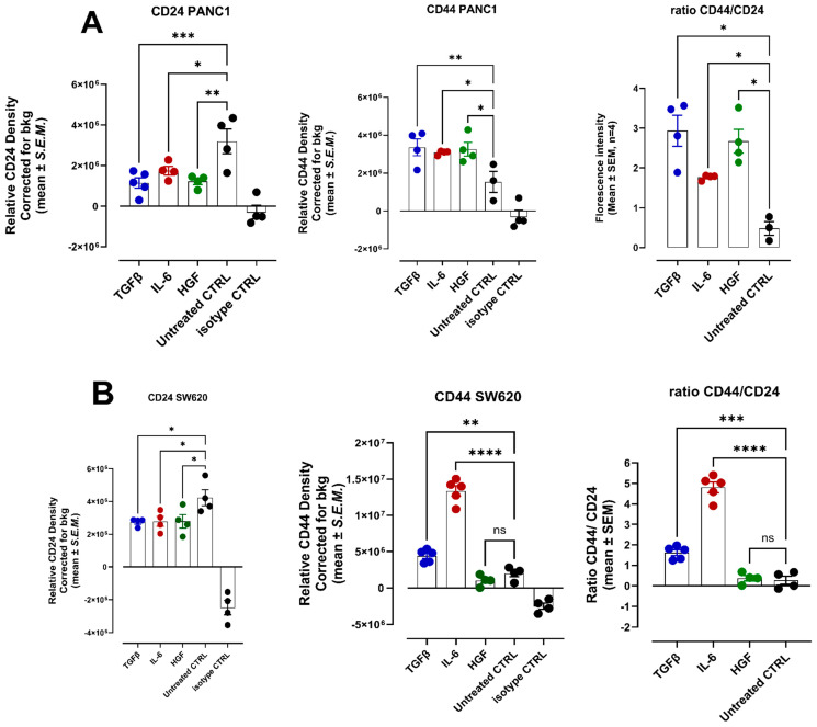
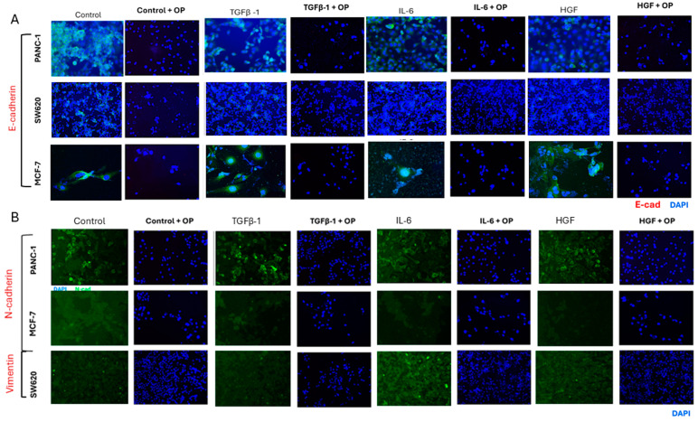
The significance of cytokine signaling on cancer progression and metastasis has raised interest in cancer research over the last few decades. Here, we analyzed the effects of three cytokines that we previously reported are significantly upregulated rapidly after the surgical removal of primary breast, colorectal, and prostate cancer. We also investigated the regulation of their cognate receptors. All experiments were conducted using the PANC-1, SW620, and MCF-7 cell lines, treated with three different cytokines (TGF-β1, HGF, and IL-6). The effect of these cytokines on the expression of epithelial-mesenchymal transition (EMT) cell surface markers and neuraminidase-1 activity was measured via fluorescent microscopy and image analysis software. The findings show that these cytokines increase the expression of mesenchymal markers while reducing epithelial markers, corresponding to the EMT process. A strong link between cytokine receptor signaling and the Neu-1-MMP-9-GPCR crosstalk was identified, suggesting that cytokine receptor binding leads to increased Neu-1 activity and subsequent signaling pathway activation. Oseltamivir phosphate (OP) prevented sialic acid hydrolysis by neuraminidase-1 (Neu-1), leading to the downregulation of these signaling cascades. In concert with the previous work revealing the role of Neu-1 in regulating other glycosylated receptors implicated in cancer cell proliferation and EMT, targeting Neu-1 may provide effective treatment against a variety of malignancies. Most significantly, the treatment of patients with specific inhibitors of Neu-1 soon after primary cancer surgery may improve our ability to cure early-stage cancer by inhibiting the EMT process and disrupting the ability of any residual cancer cell population to metastasize.

摘要

在过去几十年里,细胞因子信号传导对癌症进展和转移的重要性引发了癌症研究领域的关注。在此,我们分析了三种细胞因子的作用,我们之前报道过,在原发性乳腺癌、结直肠癌和前列腺癌手术切除后,这三种细胞因子会迅速显著上调。我们还研究了它们同源受体的调控情况。所有实验均使用PANC - 1、SW620和MCF - 7细胞系,并用三种不同的细胞因子(TGF -β1、HGF和IL - 6)进行处理。通过荧光显微镜和图像分析软件测量这些细胞因子对上皮 - 间质转化(EMT)细胞表面标志物表达和神经氨酸酶 - 1活性的影响。研究结果表明,这些细胞因子增加了间质标志物的表达,同时降低了上皮标志物的表达,这与EMT过程相对应。我们发现细胞因子受体信号传导与Neu - 1 - MMP - 9 - GPCR串扰之间存在紧密联系,这表明细胞因子受体结合会导致Neu - 1活性增加以及随后的信号通路激活。磷酸奥司他韦(OP)可阻止神经氨酸酶 - 1(Neu - 1)水解唾液酸,从而导致这些信号级联反应的下调。与之前揭示Neu - 1在调节参与癌细胞增殖和EMT的其他糖基化受体中的作用的研究一致,靶向Neu - 1可能为多种恶性肿瘤提供有效的治疗方法。最重要的是,在原发性癌症手术后不久用Neu - 1的特异性抑制剂治疗患者,可能会通过抑制EMT过程和破坏任何残留癌细胞群体的转移能力来提高我们治愈早期癌症的能力。

https://cdn.ncbi.nlm.nih.gov/pmc/blobs/6a01/11988151/672140454abf/cancers-17-01234-g014.jpg
https://cdn.ncbi.nlm.nih.gov/pmc/blobs/6a01/11988151/f9c86fe7ee9a/cancers-17-01234-g001.jpg
https://cdn.ncbi.nlm.nih.gov/pmc/blobs/6a01/11988151/4317691d04a7/cancers-17-01234-g002.jpg
https://cdn.ncbi.nlm.nih.gov/pmc/blobs/6a01/11988151/aac9662b47de/cancers-17-01234-g003.jpg
https://cdn.ncbi.nlm.nih.gov/pmc/blobs/6a01/11988151/55ae80365b34/cancers-17-01234-g004.jpg
https://cdn.ncbi.nlm.nih.gov/pmc/blobs/6a01/11988151/7ea52049510f/cancers-17-01234-g005.jpg
https://cdn.ncbi.nlm.nih.gov/pmc/blobs/6a01/11988151/9f0af1e66dc1/cancers-17-01234-g006.jpg
https://cdn.ncbi.nlm.nih.gov/pmc/blobs/6a01/11988151/578f37b37cd4/cancers-17-01234-g007.jpg
https://cdn.ncbi.nlm.nih.gov/pmc/blobs/6a01/11988151/8674d992c185/cancers-17-01234-g008.jpg
https://cdn.ncbi.nlm.nih.gov/pmc/blobs/6a01/11988151/ee2c4fb295a7/cancers-17-01234-g009.jpg
https://cdn.ncbi.nlm.nih.gov/pmc/blobs/6a01/11988151/d39eb22029f6/cancers-17-01234-g010.jpg
https://cdn.ncbi.nlm.nih.gov/pmc/blobs/6a01/11988151/7e32e00e63a3/cancers-17-01234-g011.jpg
https://cdn.ncbi.nlm.nih.gov/pmc/blobs/6a01/11988151/ced498440620/cancers-17-01234-g012.jpg
https://cdn.ncbi.nlm.nih.gov/pmc/blobs/6a01/11988151/50ea96c85c2e/cancers-17-01234-g013.jpg
https://cdn.ncbi.nlm.nih.gov/pmc/blobs/6a01/11988151/672140454abf/cancers-17-01234-g014.jpg

相似文献

1
Pro-Inflammatory Cytokines Transactivate Glycosylated Cytokine Receptors on Cancer Cells to Induce Epithelial-Mesenchymal Transition to the Metastatic Phenotype.促炎细胞因子通过反式激活癌细胞上的糖基化细胞因子受体,诱导上皮-间质转化,形成转移表型。
Cancers (Basel). 2025 Apr 5;17(7):1234. doi: 10.3390/cancers17071234.
2
Artificial and Natural Sweeteners Biased T1R2/T1R3 Taste Receptors Transactivate Glycosylated Receptors on Cancer Cells to Induce Epithelial-Mesenchymal Transition of Metastatic Phenotype.人工甜味剂和天然甜味剂偏导 T1R2/T1R3 味觉受体,使癌细胞上的糖基化受体转激活,诱导转移表型的上皮-间充质转化。
Nutrients. 2024 Jun 12;16(12):1840. doi: 10.3390/nu16121840.
3
Functional Selectivity of Cannabinoid Type 1 G Protein-Coupled Receptor Agonists in Transactivating Glycosylated Receptors on Cancer Cells to Induce Epithelial-Mesenchymal Transition Metastatic Phenotype.大麻素 1 型 G 蛋白偶联受体激动剂在激活癌细胞上糖基化受体以诱导上皮-间充质转化转移表型方面的功能选择性。
Cells. 2024 Mar 8;13(6):480. doi: 10.3390/cells13060480.
4
Metformin reverses mesenchymal phenotype of primary breast cancer cells through STAT3/NF-κB pathways.二甲双胍通过 STAT3/NF-κB 通路逆转原发性乳腺癌细胞的间充质表型。
BMC Cancer. 2019 Jul 23;19(1):728. doi: 10.1186/s12885-019-5945-1.
5
Caffeic Acid and Metformin Inhibit Invasive Phenotype Induced by TGF-β1 in C-4I and HTB-35/SiHa Human Cervical Squamous Carcinoma Cells by Acting on Different Molecular Targets.咖啡酸和二甲双胍通过作用于不同的分子靶点抑制 TGF-β1 诱导的 C-4I 和 HTB-35/SiHa 人宫颈鳞癌细胞侵袭表型。
Int J Mol Sci. 2018 Jan 16;19(1):266. doi: 10.3390/ijms19010266.
6
MHP-1 inhibits cancer metastasis and restores topotecan sensitivity via regulating epithelial-mesenchymal transition and TGF-β signaling in human breast cancer cells.MHP-1通过调节人乳腺癌细胞中的上皮-间质转化和TGF-β信号传导来抑制癌症转移并恢复拓扑替康敏感性。
Phytomedicine. 2016 Sep 15;23(10):1053-63. doi: 10.1016/j.phymed.2016.06.013. Epub 2016 Jun 21.
7
Transforming growth factor-β 1 enhances the invasiveness of breast cancer cells by inducing a Smad2-dependent epithelial-to-mesenchymal transition.转化生长因子-β1 通过诱导 Smad2 依赖性上皮间质转化增强乳腺癌细胞的侵袭性。
Oncol Rep. 2013 Jan;29(1):219-25. doi: 10.3892/or.2012.2111. Epub 2012 Oct 30.
8
TGF-beta1 induces human alveolar epithelial to mesenchymal cell transition (EMT).转化生长因子-β1诱导人肺泡上皮细胞向间充质细胞转化(EMT)。
Respir Res. 2005 Jun 9;6(1):56. doi: 10.1186/1465-9921-6-56.
9
Synthetic CB1 Cannabinoids Promote Tunneling Nanotube Communication, Cellular Migration, and Epithelial-Mesenchymal Transition in Pancreatic PANC-1 and Colorectal SW-620 Cancer Cell Lines.合成CB1大麻素促进胰腺PANC - 1和结肠直肠SW - 620癌细胞系中的隧道纳米管通讯、细胞迁移和上皮-间质转化。
Cells. 2025 Jan 7;14(2):71. doi: 10.3390/cells14020071.
10
Novel Molecular Mechanism of Aspirin and Celecoxib Targeting Mammalian Neuraminidase-1 Impedes Epidermal Growth Factor Receptor Signaling Axis and Induces Apoptosis in Pancreatic Cancer Cells.阿司匹林和塞来昔布靶向哺乳动物神经氨酸酶-1的新型分子机制抑制表皮生长因子受体信号轴并诱导胰腺癌细胞凋亡。
Drug Des Devel Ther. 2020 Oct 8;14:4149-4167. doi: 10.2147/DDDT.S264122. eCollection 2020.

本文引用的文献

1
Contemporaneous Inflammatory, Angiogenic, Fibrogenic, and Angiostatic Cytokine Profiles of the Time-to-Tumor Development by Cancer Cells to Orchestrate Tumor Neovascularization, Progression, and Metastasis.同期癌细胞向肿瘤新生血管、进展和转移的时间发展的炎症、血管生成、纤维生成和血管生成抑制细胞因子谱。
Cells. 2024 Oct 20;13(20):1739. doi: 10.3390/cells13201739.
2
ALDH1A1 as a marker for metastasis initiating cells: A mechanistic insight.ALDH1A1 作为转移起始细胞的标志物:一种机制上的见解。
Exp Cell Res. 2024 Sep 1;442(1):114213. doi: 10.1016/j.yexcr.2024.114213. Epub 2024 Aug 22.
3
Alternative splicing in EMT and TGF-β signaling during cancer progression.
癌症进展过程中 EMT 和 TGF-β 信号转导中的可变剪接。
Semin Cancer Biol. 2024 Jun;101:1-11. doi: 10.1016/j.semcancer.2024.04.001. Epub 2024 Apr 15.
4
The cytokines in tumor microenvironment: from cancer initiation-elongation-progression to metastatic outgrowth.肿瘤微环境中的细胞因子:从癌症起始-延伸-进展到转移生长。
Crit Rev Oncol Hematol. 2024 Apr;196:104311. doi: 10.1016/j.critrevonc.2024.104311. Epub 2024 Mar 3.
5
Lipids as mediators of cancer progression and metastasis.脂质作为癌症进展和转移的介质。
Nat Cancer. 2024 Jan;5(1):16-29. doi: 10.1038/s43018-023-00702-z. Epub 2024 Jan 25.
6
Contemporaneous Perioperative Inflammatory and Angiogenic Cytokine Profiles of Surgical Breast, Colorectal, and Prostate Cancer Patients: Clinical Implications.手术治疗的乳腺癌、结直肠癌和前列腺癌患者围手术期炎症和血管生成细胞因子特征:临床意义。
Cells. 2023 Dec 4;12(23):2767. doi: 10.3390/cells12232767.
7
Mutual regulation of TGFβ-induced oncogenic EMT, cell cycle progression and the DDR.转化生长因子β(TGFβ)诱导的致癌性上皮-间质转化(EMT)、细胞周期进程与DNA损伤反应(DDR)之间的相互调节
Semin Cancer Biol. 2023 Dec;97:86-103. doi: 10.1016/j.semcancer.2023.11.009. Epub 2023 Nov 28.
8
Metabolic control of cancer metastasis: role of amino acids at secondary organ sites.癌症转移的代谢控制:二级器官部位氨基酸的作用。
Oncogene. 2023 Nov;42(47):3447-3456. doi: 10.1038/s41388-023-02868-3. Epub 2023 Oct 18.
9
Mutual regulation between glycosylation and transforming growth factor-β isoforms signaling pathway.糖基化与转化生长因子-β亚型信号通路之间的相互调节。
Int J Biol Macromol. 2023 May 1;236:123818. doi: 10.1016/j.ijbiomac.2023.123818. Epub 2023 Feb 27.
10
The Significance of Cancer Stem Cells and Epithelial-Mesenchymal Transition in Metastasis and Anti-Cancer Therapy.癌症干细胞和上皮-间质转化在转移和抗癌治疗中的意义。
Int J Mol Sci. 2023 Jan 29;24(3):2555. doi: 10.3390/ijms24032555.