Madesh Mahendra, Pericherla Sruthi, Chindhalore Swapnesh
Department of Neurosurgery, East Point College of Medical Sciences and Research Hospital, Bengaluru, Karnataka, India.
J Neurosurg Case Lessons. 2025 Apr 14;9(15). doi: 10.3171/CASE24473.
Melanotic schwannoma accounts for 1% of all nerve sheath tumors. These tumors were considered benign, but recent studies have shown their malignant propensity. They pose a diagnostic challenge given the rarity of the tumor.
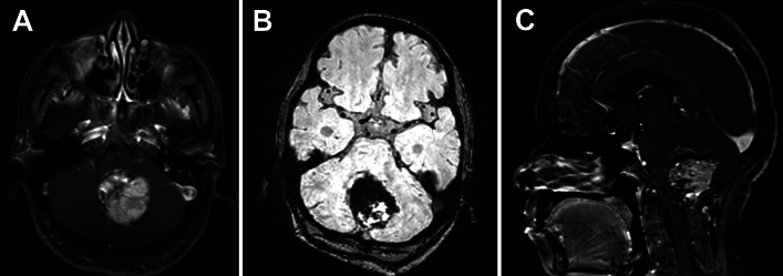
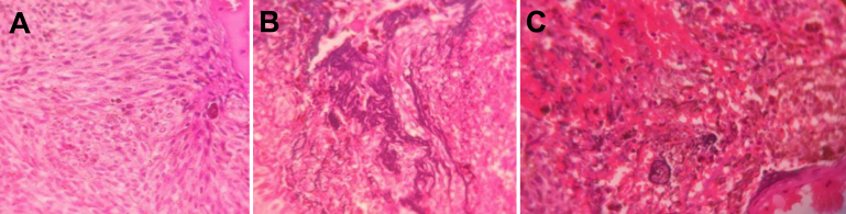
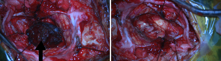
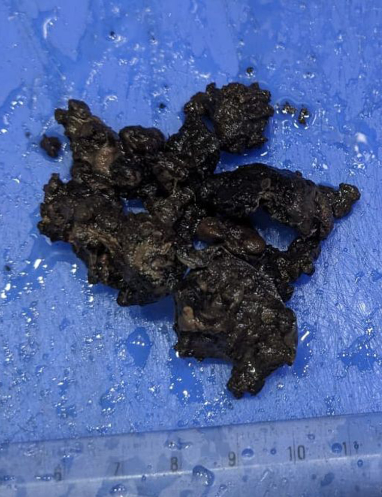
The authors report a case of a 42-year-old woman who presented with headaches and a history of frequent falls for the past year. Brain MRI revealed a lesion in the posterior fossa, effacing the median aperture and cisterna magna, which was hyperintense on T1-weighted imaging and isointense on T2-weighted imaging, with significant blooming on susceptibility-weighted imaging. The patient underwent a midline suboccipital craniotomy and gross-total resection of the tumor. Histopathological examination revealed a malignant melanotic nerve sheath tumor (MMNST) with psammoma bodies and necrosis, indicating a poor prognosis.
Melanotic schwannomas have a malignant propensity despite their benign morphology. Immunohistochemical analysis helps confirm the diagnosis of melanotic schwannoma. This is the 21st documented case of an intracranial MMNST, and, given the rarity of the tumor, there is scope for further research and studies on the role of radiotherapy in the management of these tumors. https://thejns.org/doi/10.3171/CASE24473.
黑色素性神经鞘瘤占所有神经鞘瘤的1%。这些肿瘤曾被认为是良性的,但最近的研究表明它们有恶变倾向。鉴于该肿瘤罕见,其诊断具有挑战性。
作者报告了一例42岁女性病例,该患者出现头痛,且在过去一年中有频繁跌倒史。脑部磁共振成像(MRI)显示后颅窝有一病变,压迫中脑导水管和枕大池,在T1加权成像上呈高信号,在T2加权成像上呈等信号,在磁敏感加权成像上有明显的磁敏感伪影。患者接受了枕下中线开颅手术及肿瘤全切术。组织病理学检查显示为伴有砂粒体和坏死的恶性黑色素性神经鞘瘤(MMNST),提示预后不良。
黑色素性神经鞘瘤尽管形态上为良性,但有恶变倾向。免疫组织化学分析有助于确诊黑色素性神经鞘瘤。这是第21例有文献记载的颅内MMNST病例,鉴于该肿瘤罕见,对于放疗在这些肿瘤治疗中的作用仍有进一步研究的空间。https://thejns.org/doi/10.3171/CASE24473 。