He Qinghua, Zhang Yi, Shen Mengxi, Gregori Giovanni, Rosenfeld Philip J, Wang Ruikang K
Department of Bioengineering, University of Washington, Seattle, WA, USA.
Department of Ophthalmology, Bascom Palmer Eye Institute, University of Miami Miller School of Medicine, Miami, FL, USA.
Quant Imaging Med Surg. 2025 Apr 1;15(4):2671-2681. doi: 10.21037/qims-24-2146. Epub 2025 Mar 10.
Different features of skin are associated with various medical conditions and provide opportunities to evaluate and monitor body health. This study created a strategy to assess choroidal thinning through the video analysis of facial skin.
Videos capturing the entire facial skin were collected from 48 participants with age-related macular degeneration (AMD) and 12 healthy individuals. These facial videos were analyzed using video-based trans-angiosomes imaging photoplethysmography (TaiPPG) to generate facial imaging biomarkers that were correlated with choroidal thickness (CT) measurements. The CT of all patients was determined using swept-source optical coherence tomography (SS-OCT).
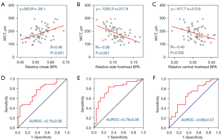
The results revealed the relationship between relative blood pulsation amplitude (BPA) in three typical facial angiosomes (cheek, side-forehead, and mid-forehead) and the average macular CT (r=0.48, P<0.001; r=-0.56, P<0.001; r=-0.40, P<0.01). When considering a diagnostic threshold of 200 µm for CT, the newly developed facial video analysis tool effectively distinguished between cases of choroidal thinning and normal cases, yielding areas under the curve of 0.75, 0.79, and 0.69.
These findings shed light on the connection between choroidal blood flow and facial skin hemodynamics, which suggests the potential for predicting vascular diseases through widely accessible skin imaging data.
皮肤的不同特征与各种医学状况相关,并为评估和监测身体健康提供了机会。本研究创建了一种通过对面部皮肤进行视频分析来评估脉络膜变薄的策略。
从48名年龄相关性黄斑变性(AMD)患者和12名健康个体中收集了捕捉整个面部皮肤的视频。使用基于视频的跨血管体成像光电容积描记法(TaiPPG)对这些面部视频进行分析,以生成与脉络膜厚度(CT)测量相关的面部成像生物标志物。所有患者的CT均使用扫频源光学相干断层扫描(SS-OCT)确定。
结果揭示了三个典型面部血管体(脸颊、前额侧面和前额中部)的相对血搏动幅度(BPA)与平均黄斑CT之间的关系(r = 0.48,P < 0.001;r = -0.56,P < 0.001;r = -0.40,P < 0.01)。当将CT的诊断阈值设定为200 µm时,新开发的面部视频分析工具能够有效区分脉络膜变薄病例和正常病例,曲线下面积分别为0.75、0.79和0.69。
这些发现揭示了脉络膜血流与面部皮肤血流动力学之间的联系,这表明通过广泛可得的皮肤成像数据预测血管疾病具有潜力。