• 文献检索
  • 文档翻译
  • 深度研究
  • 学术资讯
  • Suppr Zotero 插件Zotero 插件
  • 邀请有礼
  • 套餐&价格
  • 历史记录
应用&插件
Suppr Zotero 插件Zotero 插件浏览器插件Mac 客户端Windows 客户端微信小程序
定价
高级版会员购买积分包购买API积分包
服务
文献检索文档翻译深度研究API 文档MCP 服务
关于我们
关于 Suppr公司介绍联系我们用户协议隐私条款
关注我们

Suppr 超能文献

核心技术专利:CN118964589B侵权必究
粤ICP备2023148730 号-1Suppr @ 2026

文献检索

告别复杂PubMed语法,用中文像聊天一样搜索,搜遍4000万医学文献。AI智能推荐,让科研检索更轻松。

立即免费搜索

文件翻译

保留排版,准确专业,支持PDF/Word/PPT等文件格式,支持 12+语言互译。

免费翻译文档

深度研究

AI帮你快速写综述,25分钟生成高质量综述,智能提取关键信息,辅助科研写作。

立即免费体验

超声检查人员利用预先获取的、来自癌症扩散患病率高的特定患者样本的超声视频片段来描述卵巢癌扩散情况的能力。

Ultrasound examiners' ability to describe ovarian cancer spread using preacquired ultrasound videoclips from a selected patient sample with high prevalence of cancer spread.

作者信息

Fischerova D, Pinto P, Pesta M, Blasko M, Moruzzi M C, Testa A C, Franchi D, Chiappa V, Alcázar J L, Wiesnerova M, Cibula D, Valentin L

机构信息

Gynecologic Oncology Centre, Department of Gynecology, Obstetrics and Neonatology, First Faculty of Medicine, Charles University and General University Hospital in Prague, Prague, Czech Republic.

Department of Gynecology, Portuguese Institute of Oncology Francisco Gentil, Lisbon, Portugal.

出版信息

Ultrasound Obstet Gynecol. 2025 May;65(5):641-652. doi: 10.1002/uog.29208. Epub 2025 Apr 18.

DOI:10.1002/uog.29208
PMID:40247746
原文链接:https://pmc.ncbi.nlm.nih.gov/articles/PMC12047678/
Abstract

OBJECTIVES

To assess the ability, as well as factors affecting the ability, of ultrasound examiners with different levels of ultrasound experience to detect correctly infiltration of ovarian cancer in predefined anatomical locations, and to evaluate the inter-rater agreement regarding the presence or absence of cancer infiltration, using preacquired ultrasound videoclips obtained in a selected patient sample with a high prevalence of cancer spread.

METHODS

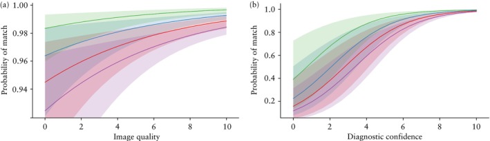
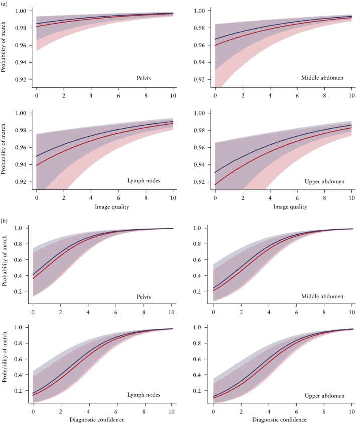
This study forms part of the Imaging Study in Advanced ovArian Cancer multicenter observational study (NCT03808792). Ultrasound videoclips showing assessment of infiltration of ovarian cancer were obtained by the principal investigator (an ultrasound expert, who did not participate in rating) at 19 predefined anatomical sites in the abdomen and pelvis, including five sites that, if infiltrated, would indicate tumor non-resectability. For each site, there were 10 videoclips showing cancer infiltration and 10 showing no cancer infiltration. The reference standard was either findings at surgery with histological confirmation or response to chemotherapy. For statistical analysis, the 19 sites were grouped into four anatomical regions: pelvis, middle abdomen, upper abdomen and lymph nodes. The videoclips were assessed by raters comprising both senior gynecologists (mainly self-trained expert ultrasound examiners who perform preoperative ultrasound assessment of ovarian cancer spread almost daily) and gynecologists who had undergone a minimum of 6 months' supervised training in the preoperative ultrasound assessment of ovarian cancer spread in a gynecological oncology center. The raters were classified as highly experienced or less experienced based on annual individual caseload and the number of years that they had been performing ultrasound evaluation of ovarian cancer spread. Raters were aware that for each site there would be 10 videoclips with and 10 without cancer infiltration. Each rater independently classified every videoclip as showing or not showing cancer infiltration and rated the image quality (on a scale from 0 to 10) and their diagnostic confidence (on a scale from 0 to 10). A generalized linear mixed model with random effects was used to estimate which factors (including level of experience, image quality, diagnostic confidence and anatomical region) affected the likelihood of a correct classification of cancer infiltration. We assessed the observed percentage of videoclips classified correctly, the expected percentage of videoclips classified correctly based on the generalized linear mixed model and inter-rater agreement (reliability) in classifying anatomical sites as being infiltrated by cancer.

RESULTS

Twenty-five raters participated in the study, of whom 13 were highly experienced and 12 were less experienced. The observed percentage of correct classification of cancer infiltration ranged from 70% to 100% depending on rater and anatomical site, and the median percentage of correct classification for the 25 raters ranged from 90% to 100%. The probability of correct classification of all 380 videoclips ranged from 0.956 to 0.975 and was not affected by the rater's level of ultrasound experience. The likelihood of correct classification increased with increased image quality and diagnostic confidence and was affected by anatomical region. It was highest for sites in the pelvis, second highest for those in the middle abdomen, third highest for lymph nodes and lowest for sites in the upper abdomen. The inter-rater agreement of all 25 raters regarding the presence of cancer infiltration ranged from substantial (Fleiss kappa, 0.68 (95% CI, 0.66-0.71)) to very good (Fleiss kappa, 0.99 (95% CI, 0.97-1.00)) depending on the anatomical site. It was lowest for sites in the upper abdomen (Fleiss kappa, 0.68 (95% CI, 0.66-0.71) to 0.97 (95% CI, 0.94-0.99)) and highest for sites in the pelvis (Fleiss kappa, 0.94 (95% CI, 0.92-0.97) to 0.99 (95% CI, 0.97-1.00)).

CONCLUSIONS

Ultrasound examiners with different levels of ultrasound experience can classify correctly predefined anatomical sites as being infiltrated or not infiltrated by ovarian cancer based on video recordings obtained by an experienced ultrasound examiner, and the inter-rater agreement is substantial. The likelihood of correct classification as well as the inter-rater agreement is highest for sites in the pelvis and lowest for sites in the upper abdomen. However, owing to the study design, our results regarding diagnostic accuracy and inter-rater agreement are likely to be overoptimistic. © 2025 The Author(s). Ultrasound in Obstetrics & Gynecology published by John Wiley & Sons Ltd on behalf of International Society of Ultrasound in Obstetrics and Gynecology.

摘要

目的

评估具有不同超声经验水平的超声检查人员正确检测卵巢癌在预定义解剖位置浸润的能力以及影响该能力的因素,并使用在癌症扩散患病率高的选定患者样本中预先获取的超声视频片段,评估检查人员之间关于癌症浸润存在与否的一致性。

方法

本研究是晚期卵巢癌影像研究多中心观察性研究(NCT03808792)的一部分。主要研究者(一位超声专家,未参与评分)在腹部和盆腔的19个预定义解剖部位获取了显示卵巢癌浸润评估的超声视频片段,其中包括5个若发生浸润则表明肿瘤不可切除的部位。对于每个部位,有10个显示癌症浸润的视频片段和10个显示无癌症浸润的视频片段。参考标准为手术发现并经组织学证实或化疗反应。为进行统计分析,将19个部位分为四个解剖区域:盆腔、中腹部、上腹部和淋巴结。视频片段由包括资深妇科医生(主要是自我训练的专家级超声检查人员,几乎每天都进行卵巢癌扩散的术前超声评估)和在妇科肿瘤中心接受过至少6个月卵巢癌扩散术前超声评估监督培训的妇科医生组成的评分者进行评估。根据每年的个人病例量以及他们进行卵巢癌扩散超声评估的年限,评分者被分为经验丰富或经验较少两类。评分者知晓每个部位会有10个有癌症浸润和10个无癌症浸润的视频片段。每位评分者独立将每个视频片段分类为显示或未显示癌症浸润,并对图像质量(从0到10评分)和他们的诊断信心(从0到10评分)进行评级。使用具有随机效应的广义线性混合模型来估计哪些因素(包括经验水平、图像质量、诊断信心和解剖区域)会影响癌症浸润正确分类的可能性。我们评估了正确分类的视频片段的观察百分比、基于广义线性混合模型正确分类的视频片段的预期百分比以及检查人员之间在将解剖部位分类为是否被癌症浸润方面的一致性(可靠性)。

结果

25名评分者参与了研究,其中13名经验丰富,12名经验较少。根据评分者和解剖部位的不同,癌症浸润正确分类的观察百分比范围为70%至100%,25名评分者正确分类中位数百分比范围为90%至100%。所有380个视频片段正确分类的概率范围为0.956至0.975,且不受评分者超声经验水平的影响。正确分类的可能性随着图像质量和诊断信心的提高而增加,并受解剖区域影响。盆腔部位最高,中腹部其次,淋巴结第三,上腹部部位最低。所有25名评分者关于癌症浸润存在与否的检查人员间一致性根据解剖部位不同,从“实质性”(Fleiss卡方值,0.68(95%CI,0.66 - 0.71))到“非常好”(Fleiss卡方值,0.99(95%CI,0.97 - 1.00))不等。上腹部部位最低(Fleiss卡方值,0.68(95%CI,0.66 - 0.71)至0.97(95%CI,0.94 - 0.99)),盆腔部位最高(Fleiss卡方值,0.94(95%CI,0.92 - 0.97)至0.99(95%CI,0.97 - 1.00))。

结论

具有不同超声经验水平的超声检查人员可以根据经验丰富的超声检查人员获取的视频记录,正确地将预定义解剖部位分类为是否被卵巢癌浸润,且检查人员间一致性较强。盆腔部位正确分类的可能性以及检查人员间一致性最高,上腹部部位最低。然而,由于研究设计的原因,我们关于诊断准确性和检查人员间一致性的结果可能过于乐观。© 2025作者。《妇产科超声》由John Wiley & Sons Ltd代表国际妇产科超声学会出版。

https://cdn.ncbi.nlm.nih.gov/pmc/blobs/c2e7/12047678/f525d8a78483/UOG-65-641-g004.jpg
https://cdn.ncbi.nlm.nih.gov/pmc/blobs/c2e7/12047678/a3b2b8a5b421/UOG-65-641-g006.jpg
https://cdn.ncbi.nlm.nih.gov/pmc/blobs/c2e7/12047678/f525d8a78483/UOG-65-641-g004.jpg
https://cdn.ncbi.nlm.nih.gov/pmc/blobs/c2e7/12047678/a3b2b8a5b421/UOG-65-641-g006.jpg
https://cdn.ncbi.nlm.nih.gov/pmc/blobs/c2e7/12047678/f525d8a78483/UOG-65-641-g004.jpg

相似文献

1
Ultrasound examiners' ability to describe ovarian cancer spread using preacquired ultrasound videoclips from a selected patient sample with high prevalence of cancer spread.超声检查人员利用预先获取的、来自癌症扩散患病率高的特定患者样本的超声视频片段来描述卵巢癌扩散情况的能力。
Ultrasound Obstet Gynecol. 2025 May;65(5):641-652. doi: 10.1002/uog.29208. Epub 2025 Apr 18.
2
Ultrasound in preoperative assessment of pelvic and abdominal spread in patients with ovarian cancer: a prospective study.超声在卵巢癌患者盆腔和腹部播散术前评估中的应用:一项前瞻性研究。
Ultrasound Obstet Gynecol. 2017 Feb;49(2):263-274. doi: 10.1002/uog.15942.
3
Preoperative staging of ovarian cancer: comparison between ultrasound, CT and whole-body diffusion-weighted MRI (ISAAC study).卵巢癌的术前分期:超声、CT与全身扩散加权磁共振成像的比较(ISAAC研究)
Ultrasound Obstet Gynecol. 2022 Feb;59(2):248-262. doi: 10.1002/uog.23654.
4
Imaging in gynecological disease (17): ultrasound features of malignant ovarian yolk sac tumors (endodermal sinus tumors).妇科疾病的影像学(17):卵巢恶性卵黄囊瘤(内胚窦瘤)的超声特征
Ultrasound Obstet Gynecol. 2020 Aug;56(2):276-284. doi: 10.1002/uog.22002.
5
The prediction of pouch of Douglas obliteration using offline analysis of the transvaginal ultrasound 'sliding sign' technique: inter- and intra-observer reproducibility.经阴道超声“滑动征”技术的离线分析预测道格拉斯窝消失:观察者间和观察者内的可重复性。
Hum Reprod. 2013 May;28(5):1237-46. doi: 10.1093/humrep/det044. Epub 2013 Mar 12.
6
Interobserver agreement of transvaginal ultrasound and magnetic resonance imaging in local staging of cervical cancer.经阴道超声与磁共振成像在宫颈癌局部分期中的观察者间一致性。
Ultrasound Obstet Gynecol. 2021 Nov;58(5):773-779. doi: 10.1002/uog.23662.
7
Intra- and interobserver agreement of proposed objective transvaginal ultrasound image-quality scoring system for use in artificial intelligence algorithm development.用于人工智能算法开发的拟议客观经阴道超声图像质量评分系统的观察者内和观察者间一致性
Ultrasound Obstet Gynecol. 2025 Mar;65(3):364-371. doi: 10.1002/uog.29178. Epub 2025 Jan 24.
8
Ultrasound diagnosis of endometrial cancer by subjective pattern recognition in women with postmenopausal bleeding: prospective inter-rater agreement and reliability study.绝经后出血妇女的主观模式识别超声诊断子宫内膜癌:前瞻性观察者间一致性和可靠性研究。
Ultrasound Obstet Gynecol. 2021 Mar;57(3):471-477. doi: 10.1002/uog.22141. Epub 2021 Feb 10.
9
International Endometrial Tumor Analysis (IETA) terminology in women with postmenopausal bleeding and sonographic endometrial thickness ≥ 4.5 mm: agreement and reliability study.绝经后出血且超声子宫内膜厚度≥4.5mm 患者的国际子宫内膜肿瘤分析(IETA)术语:一致性和可靠性研究。
Ultrasound Obstet Gynecol. 2018 Feb;51(2):259-268. doi: 10.1002/uog.18813.
10
Interobserver agreement in describing adnexal masses using the International Ovarian Tumor Analysis simple rules in a real-time setting and using three-dimensional ultrasound volumes and digital clips.在实时环境中,使用国际卵巢肿瘤分析简单规则,并利用三维超声容积和数字片段描述附件包块时的观察者间一致性。
Ultrasound Obstet Gynecol. 2014 Jul;44(1):95-9. doi: 10.1002/uog.13254. Epub 2014 May 21.

本文引用的文献

1
Staging by imaging in gynecologic cancer and the role of ultrasound: an update of European joint consensus statements.妇科癌症的影像学分期和超声的作用:欧洲联合共识声明的更新。
Int J Gynecol Cancer. 2024 Mar 4;34(3):363-378. doi: 10.1136/ijgc-2023-004609.
2
ESGO-ESMO-ESP consensus conference recommendations on ovarian cancer: pathology and molecular biology and early, advanced and recurrent disease.ESGO-ESMO-ESP 共识会议关于卵巢癌的建议:病理学和分子生物学以及早期、晚期和复发性疾病。
Ann Oncol. 2024 Mar;35(3):248-266. doi: 10.1016/j.annonc.2023.11.015. Epub 2024 Feb 1.
3
Ultrasound for assessing tumor spread in ovarian cancer. A systematic review of the literature and meta-analysis.
超声评估卵巢癌肿瘤扩散。文献系统评价和荟萃分析。
Eur J Obstet Gynecol Reprod Biol. 2024 Jan;292:194-200. doi: 10.1016/j.ejogrb.2023.11.017. Epub 2023 Nov 18.
4
Diagnostic performance of ultrasound in assessing the extension of disease in advanced ovarian cancer.超声在评估晚期卵巢癌疾病进展程度中的诊断性能。
Am J Obstet Gynecol. 2022 Oct;227(4):601.e1-601.e20. doi: 10.1016/j.ajog.2022.05.029. Epub 2022 Jun 23.
5
Interobserver agreement of transvaginal ultrasound and magnetic resonance imaging in local staging of cervical cancer.经阴道超声与磁共振成像在宫颈癌局部分期中的观察者间一致性。
Ultrasound Obstet Gynecol. 2021 Nov;58(5):773-779. doi: 10.1002/uog.23662.
6
Preoperative staging of ovarian cancer: comparison between ultrasound, CT and whole-body diffusion-weighted MRI (ISAAC study).卵巢癌的术前分期:超声、CT与全身扩散加权磁共振成像的比较(ISAAC研究)
Ultrasound Obstet Gynecol. 2022 Feb;59(2):248-262. doi: 10.1002/uog.23654.
7
Assessment of diaphragmatic function by ultrasonography: Current approach and perspectives.超声评估膈肌功能:当前方法与展望。
World J Clin Cases. 2020 Jun 26;8(12):2408-2424. doi: 10.12998/wjcc.v8.i12.2408.
8
ESMO-ESGO consensus conference recommendations on ovarian cancer: pathology and molecular biology, early and advanced stages, borderline tumours and recurrent disease.欧洲肿瘤内科学会(ESMO)-欧洲妇科肿瘤学会(ESGO)关于卵巢癌的共识会议建议:病理学与分子生物学、早期和晚期阶段、交界性肿瘤及复发性疾病
Int J Gynecol Cancer. 2019 May 7;29(4):728-760. doi: 10.1136/ijgc-2019-000308.
9
Pre-operative assessment of intra-abdominal disease spread in epithelial ovarian cancer: a comparative study between ultrasound and computed tomography.上皮性卵巢癌腹内疾病播散的术前评估:超声与计算机断层扫描的比较研究
Int J Gynecol Cancer. 2019 Feb;29(2):227-233. doi: 10.1136/ijgc-2018-000066. Epub 2019 Jan 10.
10
Ultrasound in preoperative assessment of pelvic and abdominal spread in patients with ovarian cancer: a prospective study.超声在卵巢癌患者盆腔和腹部播散术前评估中的应用:一项前瞻性研究。
Ultrasound Obstet Gynecol. 2017 Feb;49(2):263-274. doi: 10.1002/uog.15942.