• 文献检索
  • 文档翻译
  • 深度研究
  • 学术资讯
  • Suppr Zotero 插件Zotero 插件
  • 邀请有礼
  • 套餐&价格
  • 历史记录
应用&插件
Suppr Zotero 插件Zotero 插件浏览器插件Mac 客户端Windows 客户端微信小程序
定价
高级版会员购买积分包购买API积分包
服务
文献检索文档翻译深度研究API 文档MCP 服务
关于我们
关于 Suppr公司介绍联系我们用户协议隐私条款
关注我们

Suppr 超能文献

核心技术专利:CN118964589B侵权必究
粤ICP备2023148730 号-1Suppr @ 2026

文献检索

告别复杂PubMed语法,用中文像聊天一样搜索,搜遍4000万医学文献。AI智能推荐,让科研检索更轻松。

立即免费搜索

文件翻译

保留排版,准确专业,支持PDF/Word/PPT等文件格式,支持 12+语言互译。

免费翻译文档

深度研究

AI帮你快速写综述,25分钟生成高质量综述,智能提取关键信息,辅助科研写作。

立即免费体验

肝硬化性血管瘤的诊断陷阱:一例报告

Diagnostic Pitfalls of Hepatic Sclerosed Hemangiomas: A Case Report.

作者信息

Takihara Hiroshi, Oura Shoji, Shintani Hiroshi, Tanaka Hiroto

机构信息

Department of Gastroenterology, Uji Tokushukai Hospital, Uji, JPN.

Department of Gastroenterology, Kishiwada Tokushukai Hospital, Kishiwada, JPN.

出版信息

Cureus. 2025 Mar 17;17(3):e80738. doi: 10.7759/cureus.80738. eCollection 2025 Mar.

DOI:10.7759/cureus.80738
PMID:40248561
原文链接:https://pmc.ncbi.nlm.nih.gov/articles/PMC12003112/
Abstract

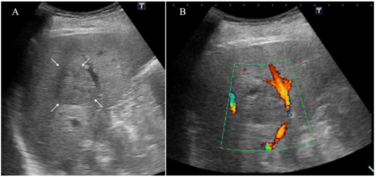
An 81-year-old man was found to have a liver mass on an annual medical checkup. Enhanced CT of the mass, 3.8 cm in size, showed weak enhancement with a small non-enhanced oval area near the mass borders. Ultrasound showed an oval mass with internal iso-echoes. Magnetic resonance imaging (MRI) showed that the mass had low and slightly high signal intensities on T1- and T2-weighted images, respectively. MRI of the small non-enhanced area on CT showed high signal intensity both on T1- and T2-weighted images, suggesting focal subacute bleeding. In addition to these image findings, elevated serum α-fetoprotein (AFP) and lectin-reactive fraction of AFP levels made us resect the liver mass without performing a biopsy to the tumor under the tentative diagnosis of possible hepatic malignancy. A postoperative pathological study showed that the mass had massive scar tissue with hemorrhage, lymphocytes, hemosiderin-laden macrophages, and multiple vascular structures, leading to the diagnosis of a hepatic sclerosed hemangioma (HSH). Why this case showed high tumor marker levels remains uncertain. The patient showed normal tumor marker levels shortly after surgery and has been well for 40 months without any problems. Diagnostic physicians should note that HSHs can present very similar image findings to those of intra-hepatic cholangiocarcinomas.

摘要

一名81岁男性在年度体检中被发现肝脏有肿物。该肿物大小为3.8 cm,增强CT显示强化较弱,肿物边界附近有一小片无强化的椭圆形区域。超声显示为椭圆形肿物,内部回声均匀。磁共振成像(MRI)显示,该肿物在T1加权像上呈低信号,在T2加权像上呈稍高信号。CT上无强化的小区域的MRI在T1加权像和T2加权像上均显示高信号,提示局灶性亚急性出血。除了这些影像学表现外,血清甲胎蛋白(AFP)升高以及AFP的凝集素反应性部分水平升高,使我们在初步诊断为可能的肝脏恶性肿瘤的情况下,未对肿瘤进行活检就切除了肝脏肿物。术后病理研究显示,肿物有大量瘢痕组织,伴有出血、淋巴细胞、含铁血黄素巨噬细胞和多个血管结构,诊断为肝脏硬化性血管瘤(HSH)。该病例为何出现肿瘤标志物水平升高仍不确定。患者术后不久肿瘤标志物水平恢复正常,40个月来情况良好,无任何问题。诊断医生应注意,HSH的影像学表现可能与肝内胆管癌非常相似。

https://cdn.ncbi.nlm.nih.gov/pmc/blobs/e3e5/12003112/71fbb4f4e971/cureus-0017-00000080738-i04.jpg
https://cdn.ncbi.nlm.nih.gov/pmc/blobs/e3e5/12003112/930d15d53094/cureus-0017-00000080738-i01.jpg
https://cdn.ncbi.nlm.nih.gov/pmc/blobs/e3e5/12003112/273a05010a63/cureus-0017-00000080738-i02.jpg
https://cdn.ncbi.nlm.nih.gov/pmc/blobs/e3e5/12003112/624d3c51b1e0/cureus-0017-00000080738-i03.jpg
https://cdn.ncbi.nlm.nih.gov/pmc/blobs/e3e5/12003112/71fbb4f4e971/cureus-0017-00000080738-i04.jpg

相似文献

1
Diagnostic Pitfalls of Hepatic Sclerosed Hemangiomas: A Case Report.肝硬化性血管瘤的诊断陷阱:一例报告
Cureus. 2025 Mar 17;17(3):e80738. doi: 10.7759/cureus.80738. eCollection 2025 Mar.
2
Hepatic sclerosed hemangioma and sclerosing cavernous hemangioma: a radiological study.肝硬化性血管瘤和硬化性海绵状血管瘤:影像学研究。
Jpn J Radiol. 2021 Nov;39(11):1059-1068. doi: 10.1007/s11604-021-01139-z. Epub 2021 May 27.
3
Preoperative diagnosis of the sclerosed hemangioma of the liver using multimodality imaging findings: A case report.利用多模态影像学表现对肝脏硬化性血管瘤进行术前诊断:病例报告
Radiol Case Rep. 2018 Jun 1;13(5):1025-1029. doi: 10.1016/j.radcr.2018.04.007. eCollection 2018 Oct.
4
A hepatic sclerosed hemangioma with significant morphological change over a period of 10 years: a case report.10年间形态发生显著变化的肝脏硬化性血管瘤1例报告
J Med Case Rep. 2013 May 28;7:139. doi: 10.1186/1752-1947-7-139.
5
Differentiation of Hepatic Sclerosed Hemangiomas From Cavernous Hemangiomas Based on Gadoxetic Acid-Enhanced Magnetic Resonance Imaging Features.基于钆塞酸增强磁共振成像特征鉴别肝硬化性血管瘤与海绵状血管瘤
J Comput Assist Tomogr. 2019 Sep/Oct;43(5):762-769. doi: 10.1097/RCT.0000000000000905.
6
Hepatic Sclerosed Hemangioma: a case report and review of the literature.肝硬化性血管瘤:一例病例报告及文献复习
BMC Surg. 2015 Apr 17;15:45. doi: 10.1186/s12893-015-0029-x.
7
Hepatic Sclerosing Hemangioma with Predominance of the Sclerosed Area Mimicking a Biliary Cystadenocarcinoma.以硬化区域为主的肝脏硬化性血管瘤,酷似胆管囊腺癌。
Case Reports Hepatol. 2018 Oct 4;2018:7353170. doi: 10.1155/2018/7353170. eCollection 2018.
8
[Multiple hepatic sclerosed hemangiomas that are difficult to diagnose preoperatively:a case report].[术前难以诊断的多发性肝脏硬化性血管瘤:1例报告]
Nihon Shokakibyo Gakkai Zasshi. 2018;115(5):485-493. doi: 10.11405/nisshoshi.115.485.
9
Sclerosed hemangioma and sclerosing cavernous hemangioma of the liver: a comparative clinicopathologic and immunohistochemical study with emphasis on the role of mast cells in their histogenesis.肝脏硬化性血管瘤和硬化性海绵状血管瘤:一项比较临床病理和免疫组化研究,重点关注肥大细胞在其组织发生中的作用
Liver. 2002 Feb;22(1):70-8. doi: 10.1046/j.0106-9543.2001.01604.x.
10
A hepatic sclerosed hemangioma with drastic changes in contrast-enhanced ultrasonography.肝硬化性血管瘤,超声造影有显著变化。
Clin J Gastroenterol. 2020 Dec;13(6):1252-1257. doi: 10.1007/s12328-020-01194-5. Epub 2020 Jul 23.

引用本文的文献

1
[Vascular neoplasms of the liver].[肝脏血管肿瘤]
Pathologie (Heidelb). 2025 Sep 15. doi: 10.1007/s00292-025-01468-0.

本文引用的文献

1
Posterior Echo Enhancement by Elastosis in Breast Cancer: A Case Report.乳腺癌中弹性组织变性所致后方回声增强:一例报告
Cureus. 2025 Jan 26;17(1):e78034. doi: 10.7759/cureus.78034. eCollection 2025 Jan.
2
Low Fluorodeoxyglucose Uptake in the Metastatic Lung Tumor From Clear Cell Renal Cell Carcinoma.透明细胞肾细胞癌肺转移瘤中低氟脱氧葡萄糖摄取情况
Cureus. 2024 Aug 26;16(8):e67854. doi: 10.7759/cureus.67854. eCollection 2024 Aug.
3
Hepatic sclerosed hemangioma and sclerosing cavernous hemangioma: a radiological study.肝硬化性血管瘤和硬化性海绵状血管瘤:影像学研究。
Jpn J Radiol. 2021 Nov;39(11):1059-1068. doi: 10.1007/s11604-021-01139-z. Epub 2021 May 27.
4
Resection as first-line therapy for large hepatic sclerosing hemangioma: a case report.肝巨大硬化性血管瘤的一线治疗——手术切除:一例报告
Onco Targets Ther. 2019 Aug 22;12:6839-6842. doi: 10.2147/OTT.S217528. eCollection 2019.
5
Clinical case report: Sclerosing hemangioma of the liver, a rare but great mimicker.临床病例报告:肝脏硬化性血管瘤,一种罕见但极具迷惑性的病症。
Radiol Case Rep. 2016 May 6;11(2):58-61. doi: 10.1016/j.radcr.2016.02.015. eCollection 2016 Jun.
6
Hepatic Sclerosed Hemangioma: a case report and review of the literature.肝硬化性血管瘤:一例病例报告及文献复习
BMC Surg. 2015 Apr 17;15:45. doi: 10.1186/s12893-015-0029-x.
7
Multiple hepatic sclerosing hemangioma mimicking metastatic liver tumor successfully treated by laparoscopic surgery: Report of a case.腹腔镜手术成功治疗酷似肝转移瘤的多发性硬化性肝血管瘤:病例报告
Int J Surg Case Rep. 2015;8C:137-40. doi: 10.1016/j.ijscr.2015.01.032. Epub 2015 Jan 21.
8
A sclerosing hemangioma of the liver.肝脏硬化性血管瘤
Clin Mol Hepatol. 2013 Dec;19(4):426-30. doi: 10.3350/cmh.2013.19.4.426.
9
Hepatic haemangioma: common and uncommon imaging features.肝血管瘤:常见和不常见的影像学特征。
Diagn Interv Imaging. 2013 Sep;94(9):849-59. doi: 10.1016/j.diii.2013.04.008. Epub 2013 Jun 21.
10
Sclerosing hemangioma in the liver.肝脏硬化性血管瘤
Korean J Hepatol. 2011 Sep;17(3):242-6. doi: 10.3350/kjhep.2011.17.3.242.