Zhou Linyi, Tao Kejin, Ma Jinjin, Pan Xianglong, Zhang Kedie, Feng Jianying
School/Hospital of Stomatology, Zhejiang Chinese Medical University, Hangzhou, Zhejiang, 310053, China.
Sir Run Run Shaw Hospital Medical School ZheJiang University, Hangzhou, Zhejiang, 310016, China.
BMC Oral Health. 2025 Apr 21;25(1):611. doi: 10.1186/s12903-025-05991-7.
Analyse the correlation between the changes in joint space of TMJ and the displacement and degree of articular disc for clinical diagnosis.
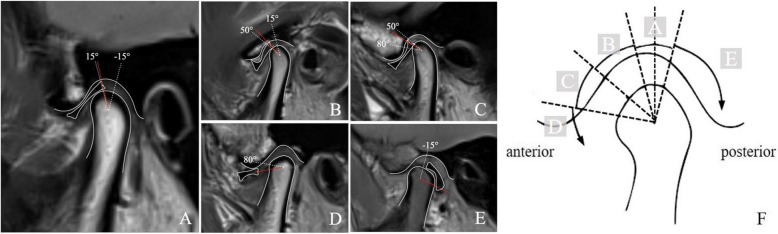
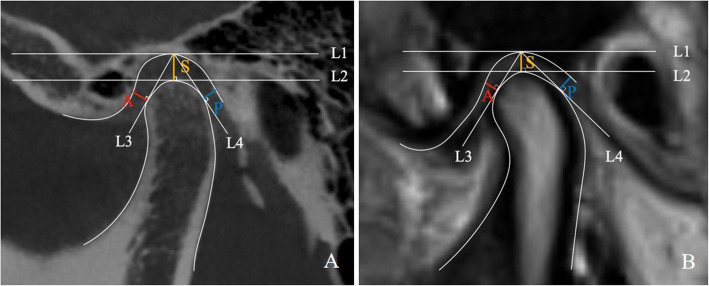
Two hundred sixteen TMJs of 108 temporomandibular disorders (TMD) patients with clinical symptoms and MRI examination were included in the study. 30 of these patients had undergone CBCT before MRI. According to the degree of articular disc displacement, the 216 joints are divided into five groups. Group A: no disc displacement (40 cases); group B: mild anterior disc displacement (44 cases); group C: moderate anterior disc displacement (36 cases); group D: severe anterior disc displacement (52 cases); group E: posterior displacement (44 cases). The 132 sides of these anteriorly displaced discs (ADD) were further divided into two groups, anterior disc displacement with reduction (ADDwR) and anterior disc displacement without reduction (ADDwoR). We analysed the concordance of the joint space measured by MRI and CBCT, and explored the relationship between joint space, ln(P/A) values and joint disc displacement.
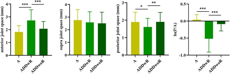
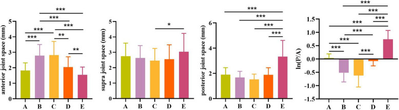
There was no statistically significant difference between the joint spaces measured by CBCT and MRI (P > 0.05). The anterior joint space in group B (2.7 ± 0.72 mm) and C (2.82 ± 0.88 mm) was larger than group A (1.82 ± 0.50 mm) (P < 0.05), and ln(P/A) value in group B (-0.52 ± 0.34) and C (-0.62 ± 0.43) was smaller than group A (0.04 ± 0.15) (P < 0.05). The posterior joint space (3.33 ± 1.28 mm) and ln(P/A) value (0.74 ± 0.33) in group E was larger than group A (P < 0.05). There was no significant difference in the anterior, superior and posterior joint space and ln(P/A) value between group D and A (P > 0.05). The ADDwR group had a larger anterior joint space (2.72 ± 0.83 mm) than group A (P < 0.05), while having a smaller posterior joint space (1.61 ± 0.49 mm) and ln(P/A) value (-0.52 ± 0.39 mm) (P < 0.05). Compared with group A, there was no significant difference in the anterior joint space and ln(P/A) value in the ADDwoR group(P > 0.05).
There is no significant change in anterior, supra, and posterior joint space in severe anterior disc displacement. The anterior joint space increases in mild to moderate anterior disc displacement, but does not change in severe anterior disc displacement-the posterior joint space increases when the joint disc is displaced posteriorly. The position of the joint disc cannot be accurately inferred by observing the joint space through CBCT, and a combination of MRI and clinical examination is required to make a definitive judgement.
分析颞下颌关节(TMJ)关节间隙变化与关节盘移位及程度之间的相关性,以用于临床诊断。
本研究纳入了108例有临床症状且进行了MRI检查的颞下颌关节紊乱病(TMD)患者的216个TMJ。其中30例患者在MRI检查前已接受CBCT检查。根据关节盘移位程度,将216个关节分为五组。A组:无盘移位(40例);B组:轻度盘前移位(44例);C组:中度盘前移位(36例);D组:重度盘前移位(52例);E组:盘后移位(44例)。将这些前移位盘(ADD)的132侧进一步分为两组,可复性盘前移位(ADDwR)和不可复性盘前移位(ADDwoR)。我们分析了MRI和CBCT测量的关节间隙的一致性,并探讨了关节间隙、ln(P/A)值与关节盘移位之间的关系。
CBCT和MRI测量的关节间隙之间无统计学显著差异(P>0.05)。B组(2.7±0.72mm)和C组(2.82±0.88mm)的前关节间隙大于A组(1.82±0.50mm)(P<0.05),B组(-0.52±0.34)和C组(-0.62±0.43)的ln(P/A)值小于A组(0.04±0.15)(P<0.05)。E组的后关节间隙(3.33±1.28mm)和ln(P/A)值(0.74±0.33)大于A组(P<0.05)。D组与A组在前、上、后关节间隙及ln(P/A)值方面无显著差异(P>0.05)。ADDwR组的前关节间隙(2.72±0.83mm)大于A组(P<0.05),而后关节间隙(1.61±0.49mm)和ln(P/A)值(-0.52±0.39mm)较小(P<0.05)。与A组相比,ADDwoR组的前关节间隙和ln(P/A)值无显著差异(P>0.05)。
重度盘前移位时,前、上、后关节间隙无显著变化。轻度至中度盘前移位时前关节间隙增大,但重度盘前移位时不变——关节盘向后移位时后关节间隙增大。通过CBCT观察关节间隙无法准确推断关节盘的位置,需要结合MRI和临床检查才能做出明确判断。