Shen Jiaqi, Liu Xin, Lin Mingyue, Shao Yuting, Niu Guozhen, Qu Shen, Niu Yunli, Zhou Qi, Zhang Li, Bi Yanlong
Department of Ophthalmology, Tongji Hospital, School of Medicine, Tongji University, Shanghai, China.
Department of Ophthalmology, Guizhou Provincial People's Hospital, Guiyang, China.
Front Med (Lausanne). 2025 Apr 10;12:1533950. doi: 10.3389/fmed.2025.1533950. eCollection 2025.
This study aimed to assess corneal endothelial cell loss (ECL) following phacoemulsification and intraocular lens implantation (Phaco+IOL) in eyes with acute primary angle-closure glaucoma (APACG) and cataracts under different preoperative intraocular pressure (IOP) levels.
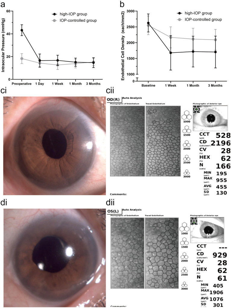
This non-randomized controlled trial included 75 eyes from 75 patients with APACG and cataracts who underwent Phaco+IOL. All patients received pharmacotherapy and anterior chamber paracentesis before surgery and were grouped according to their preoperative IOP: the high-IOP group (IOP ≥ 25 mmHg) and the IOP-controlled group (IOP < 25 mmHg). IOP, visual outcome, endothelial cell density (ECD), hexagonality (HEX), coefficient of variation (CV), and central corneal thickness (CCT) were evaluated up to 3 months postoperatively. Baseline ECD, HEX, and CV parameters were measured in the contralateral eyes of all patients as a reference and compared with the postoperative results.
The average IOP decreased from 43.2 ± 4.8 mmHg to 16.5 ± 5.8 mmHg ( < 0.001) in the high-IOP group and from 18.3 ± 4.3 mmHg to 14.3 ± 3.2 mmHg ( < 0.001) in the IOP-controlled group on the first postoperative day. The changes in IOP were more significant in the high-IOP group ( < 0.001). The ECD at 3 months was 1705.2 ± 503.8 cells/mm in the high-IOP group and 2091.8 ± 330.1 cells/mm in the IOP-controlled group ( < 0.001). The ECL rates at 3 months were 35.0% (high-IOP group) and 17.4% (IOP-controlled group) ( < 0.001). The postoperative changes in HEX and CV at 3 months were more significant in the high-IOP group ( < 0.001; = 0.003). Both groups produced comparable improvements in visual acuity and IOP.
Uncontrolled high IOP (≥ 25 mmHg) before Phaco+IOL in patients with APACG and cataracts is associated with a higher rate of ECL. The rapid and substantial reduction of IOP during surgery may exacerbate corneal endothelial cell loss.
ClinicalTrails.gov, identifier ChiCTR2100052096.
本研究旨在评估急性原发性闭角型青光眼(APACG)合并白内障患者在不同术前眼压(IOP)水平下,行超声乳化白内障吸除联合人工晶状体植入术(Phaco+IOL)后的角膜内皮细胞丢失(ECL)情况。
本非随机对照试验纳入了75例接受Phaco+IOL的APACG合并白内障患者的75只眼。所有患者在手术前均接受药物治疗和前房穿刺,并根据术前眼压分组:高眼压组(IOP≥25mmHg)和眼压控制组(IOP<25mmHg)。术后3个月内评估眼压、视力、内皮细胞密度(ECD)、六角形细胞比例(HEX)、变异系数(CV)和中央角膜厚度(CCT)。测量所有患者对侧眼的基线ECD、HEX和CV参数作为参考,并与术后结果进行比较。
术后第1天,高眼压组平均眼压从43.2±4.8mmHg降至16.5±5.8mmHg(<0.001),眼压控制组从18.3±4.3mmHg降至14.3±3.2mmHg(<0.001)。高眼压组眼压变化更显著(<0.001)。高眼压组术后3个月ECD为1705.2±503.8个细胞/mm,眼压控制组为2091.8±330.1个细胞/mm(<0.001)。术后3个月ECL率分别为35.0%(高眼压组)和17.4%(眼压控制组)(<0.001)。高眼压组术后3个月HEX和CV的变化更显著(<0.001;=0.003)。两组在视力和眼压改善方面相当。
APACG合并白内障患者在Phaco+IOL术前眼压未控制(≥25mmHg)与较高的ECL率相关。手术中眼压的快速大幅降低可能会加剧角膜内皮细胞丢失。
ClinicalTrails.gov,标识符ChiCTR2100052096。