Markovic Ksenija, Djuricic Goran, Milojkovic Djordje, Banovac Dusan, Davidovic Kristina, Vasin Dragan, Sisevic Jelena, Zagorac Slavisa, Gluscevic Boris, Bokonjic Dejan, Djulejic Vuk, Milic Natasa
Institute for Medical Statistics and Informatics, Faculty of Medicine, University of Belgrade, 11000 Belgrade, Serbia.
Department of Diagnostic Imaging, University Children's Hospital, 11000 Belgrade, Serbia.
J Clin Med. 2025 Apr 16;14(8):2728. doi: 10.3390/jcm14082728.
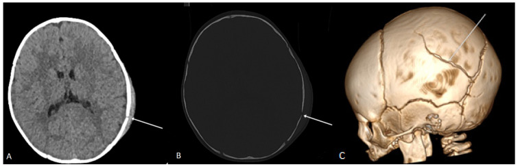
: Mild traumatic brain injury (mTBI) is a leading cause of pediatric emergency department visits, particularly among children under three years old. Although computed tomography (CT) is the gold standard for diagnosing intracranial injuries, its use in young children poses radiation risks. Identifying reliable clinical indicators that justify CT imaging is essential for optimizing both patient safety and resource utilization. Objective: This study aimed to evaluate CT findings in children under three years of age with mTBI and no focal neurological deficits, as well as to identify clinical predictors associated with skull fractures and intracranial injuries. : A retrospective analysis was conducted on 224 children under 36 months who presented with mTBI to a tertiary pediatric hospital from July 2019 to July 2024. Demographic data, injury mechanisms, clinical presentation and CT findings were evaluated. Univariate and multivariate regression analyses were performed to identify risk factors associated with skull fractures and intracranial injuries. : Falls accounted for 96.4% of injuries, with the majority occurring from heights of 0.5-1 m. The parietal region was the most frequently affected site (38%). Skull fractures were present in 46% of cases and were primarily linear (92.8%). Intracranial hematomas were identified in 13.8% of cases, while brain edema was observed in 7.6%. Significant predictors of skull fractures included age under 12 months ( < 0.001), falls from 0.5-1 m ( = 0.005), somnolence ( = 0.030), scalp swelling ( = 0.001) and indentation of the scalp ( = 0.016). Parietal bone involvement was the strongest predictor of both skull fractures (OR = 7.116, < 0.001) and intracranial hematomas (OR = 4.993, < 0.001). Conversely, frontal bone involvement was associated with a lower likelihood of fractures and hematomas. The findings highlight key clinical indicators that can guide decision-making for CT imaging in children with mTBI. Infants under 12 months, falls from moderate heights and parietal bone involvement significantly increase the risk of fractures and intracranial injuries. A more refined diagnostic approach could help reduce unnecessary CT scans while ensuring the timely identification of clinically significant injuries.
轻度创伤性脑损伤(mTBI)是儿科急诊科就诊的主要原因,尤其是在三岁以下儿童中。尽管计算机断层扫描(CT)是诊断颅内损伤的金标准,但其在幼儿中的使用存在辐射风险。确定可靠的临床指标以证明CT成像的合理性对于优化患者安全和资源利用至关重要。目的:本研究旨在评估三岁以下无局灶性神经功能缺损的mTBI儿童的CT检查结果,并确定与颅骨骨折和颅内损伤相关的临床预测因素。:对2019年7月至2024年7月在一家三级儿科医院就诊的224名36个月以下的mTBI儿童进行了回顾性分析。评估了人口统计学数据、损伤机制、临床表现和CT检查结果。进行单因素和多因素回归分析以确定与颅骨骨折和颅内损伤相关的危险因素。:跌倒占损伤的96.4%,大多数发生在0.5-1米的高度。顶叶区域是最常受影响的部位(38%)。46%的病例存在颅骨骨折,主要为线性骨折(92.8%)。13.8%的病例发现颅内血肿,7.6%观察到脑水肿。颅骨骨折的重要预测因素包括12个月以下年龄(<0.001)、从0.5-1米高处跌倒(=0.005)、嗜睡(=0.030)、头皮肿胀(=0.001)和头皮凹陷(=0.016)。顶骨受累是颅骨骨折(OR = 7.116,<0.001)和颅内血肿(OR = 4.993,<0.001)的最强预测因素。相反,额骨受累与骨折和血肿的可能性较低相关。研究结果突出了关键的临床指标,可指导mTBI儿童CT成像的决策。12个月以下的婴儿、中等高度跌倒和顶骨受累显著增加骨折和颅内损伤的风险。一种更精细的诊断方法有助于减少不必要的CT扫描,同时确保及时识别具有临床意义的损伤。