Liu Zhao, Liu Xi, Zhou Yi, Wen Xin, Xu Jie, He Meizhi, Chen Jiongcheng, Jia Nan, Liu Youhua
State Key Laboratory of Multi-organ Injury Prevention and Treatment, Division of Nephrology, Nanfang Hospital, Southern Medical University, Guangzhou, China.
National Clinical Research Center of Kidney Disease, Guangdong Provincial Institute of Nephrology, Guangzhou, China.
Theranostics. 2025 Apr 9;15(11):5121-5137. doi: 10.7150/thno.110034. eCollection 2025.
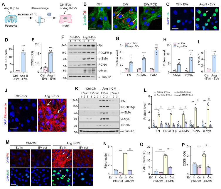
Podocyte injury leading to proteinuria is the primary feature of a vast majority of glomerular diseases, while mesangial cell activation is the hallmark of glomerulosclerosis. Whether and how these two events are connected remains elusive. In this study, we investigated the role of extracellular vesicles (EVs) in linking podocyte injury to mesangial activation in glomerular disease. EVs were characterized by nanoparticle tracking analysis and electron microscopy. Differentially expressed proteins from podocyte-derived EVs were analyzed by protein microarray. The role and mechanism by which EVs-packaged sonic hedgehog (Shh) mediates mesangial cell activation were investigated and . An increased production of EVs in mouse podocytes (MPC5) was observed after injury induced by angiotensin II (Ang II). Shh and N-Shh were identified as major constituents of the proteins encapsulated in EVs isolated from Ang II-treated MPC5 cells (Ang II-EVs). In vitro, Ang II-EVs induced the activation and proliferation of rat mesangial cells (HBZY-1), whereas inhibition of EV secretion with dimethyl amiloride, depletion of EVs from conditioned media or knockdown of Shh expression abolished the ability of Ang II-EVs to induce HBZY-1 activation. , intravenous injection of Ang II-EVs exacerbated glomerulosclerosis, which was negated by hedgehog inhibitor. Furthermore, blocking EV secretion also ameliorated glomerulosclerosis in mouse model of glomerular disease. These findings suggest that podocyte injury can cause mesangial cell activation and glomerulosclerosis by releasing Shh-enriched EVs. Therefore, strategies targeting EVs may be a novel way to ameliorate proteinuric kidney disease.
足细胞损伤导致蛋白尿是绝大多数肾小球疾病的主要特征,而系膜细胞活化是肾小球硬化的标志。这两个事件是否以及如何相互关联仍不清楚。在本研究中,我们调查了细胞外囊泡(EVs)在肾小球疾病中将足细胞损伤与系膜活化联系起来的作用。通过纳米颗粒跟踪分析和电子显微镜对EVs进行表征。通过蛋白质微阵列分析来自足细胞衍生的EVs中差异表达的蛋白质。研究了EVs包裹的音猬因子(Shh)介导系膜细胞活化的作用和机制。在用血管紧张素II(Ang II)诱导损伤后,观察到小鼠足细胞(MPC5)中EVs的产生增加。Shh和N-Shh被鉴定为从Ang II处理的MPC5细胞(Ang II-EVs)中分离出的包裹在EVs中的蛋白质的主要成分。在体外,Ang II-EVs诱导大鼠系膜细胞(HBZY-1)的活化和增殖,而用二甲基阿米洛利抑制EV分泌、从条件培养基中去除EVs或敲低Shh表达则消除了Ang II-EVs诱导HBZY-1活化的能力。此外,静脉注射Ang II-EVs加剧了肾小球硬化,而刺猬因子抑制剂可使其减轻。此外,在肾小球疾病小鼠模型中阻断EV分泌也改善了肾小球硬化。这些发现表明,足细胞损伤可通过释放富含Shh的EVs导致系膜细胞活化和肾小球硬化。因此,针对EVs的策略可能是改善蛋白尿性肾病的一种新方法。