Sun Yuying, Zhang Jieying, Wang Yilin, Zhang Xinxin, Chen Yan
Radiology Department, National Cancer Center/National Clinical Research Center for Cancer/Cancer Hospital, Chinese Academy of Medical Sciences and Peking Union Medical College, Beijing, China.
Front Oncol. 2025 Apr 15;15:1565152. doi: 10.3389/fonc.2025.1565152. eCollection 2025.
To investigate the utility of multi-sequence magnetic resonance imaging (MRI) and whole-tumor apparent diffusion coefficient (ADC) histogram metrics in preoperatively differentiating p53 abnormal (p53abn) from non-p53abn endometrial carcinoma (EC).
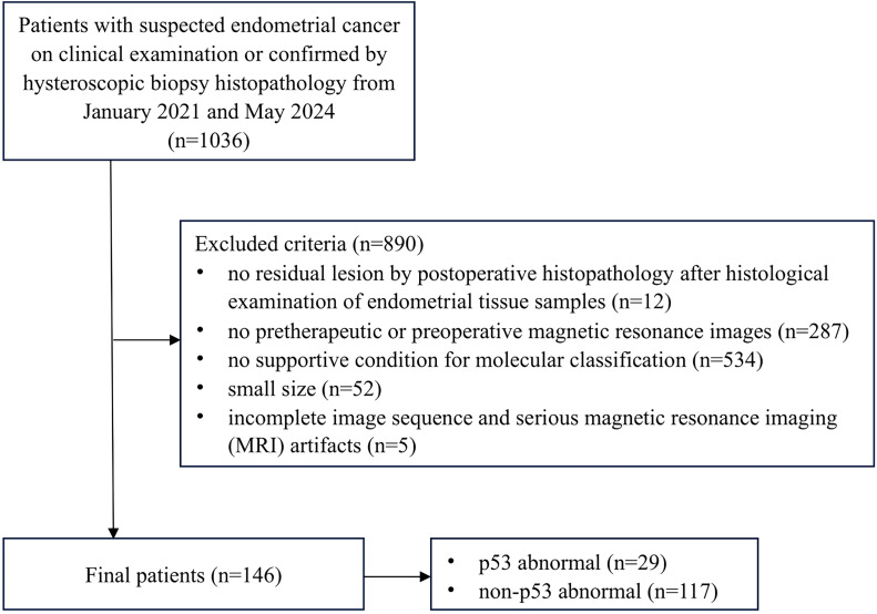
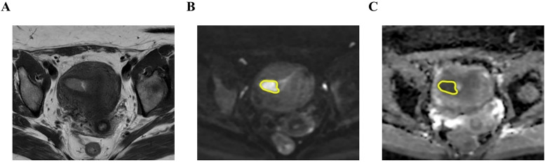
This retrospective study included 146 EC patients (29 p53abn cases and 117 non-p53abn cases) who underwent preoperative MRI scans. MRI features were analyzed. Whole-tumor ADC histogram analysis was conducted by delineating regions of interest (ROIs) on diffusion-weighted imaging (DWI) scans. Receiver operating characteristic (ROC) curve analysis with the area under the curve (AUC) was used for diagnostic performance evaluation.
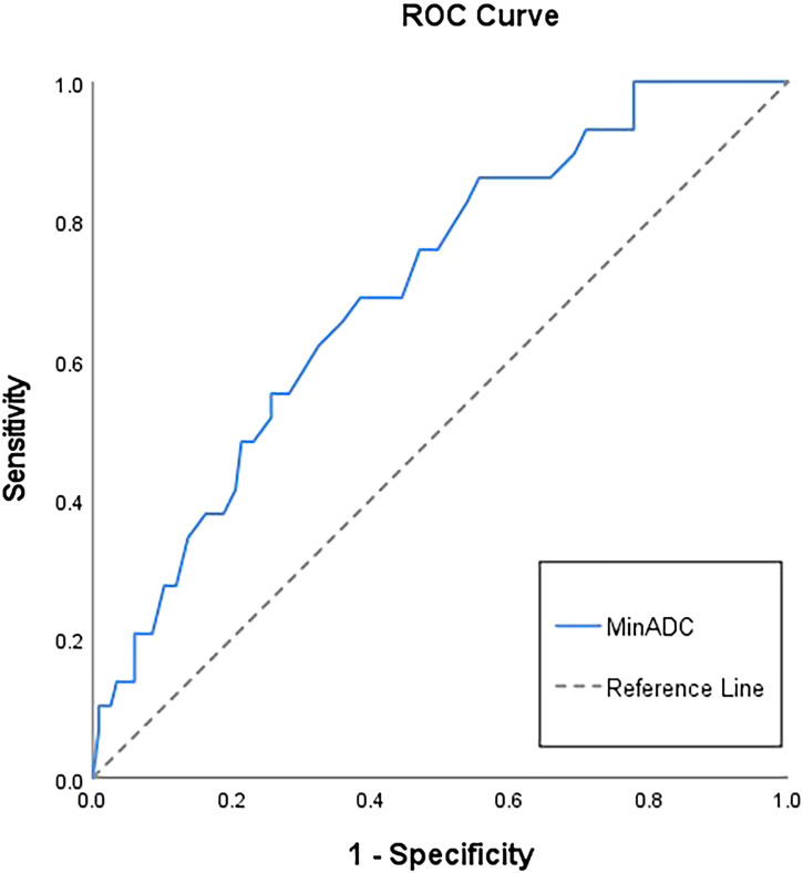
Extrauterine extension (p=0.004) and lymphadenopathy (p=0.005) were more frequently observed in p53abn EC compared to non-p53abn EC. p53abn EC exhibited significantly lower value of minADC (p=0.001), meanADC (p=0.005), P10 (p=0.009), P50 (p=0.007), and P90 (p=0.013) ADC and higher value of kurtosis (p=0.008), compared to non-p53abn EC. MinADC demonstrated the highest discrimination ability in differentiating p53abn from non-p53abn EC [AUC 0.70(0.60;0.80)].
Preoperative multi-sequence MRI findings and whole-tumor ADC histogram metrics are conducive to differentiating p53abn from non-p53abn EC.
探讨多序列磁共振成像(MRI)及全肿瘤表观扩散系数(ADC)直方图指标在术前鉴别p53异常(p53abn)与非p53abn子宫内膜癌(EC)中的应用价值。
本回顾性研究纳入了146例术前行MRI扫描的EC患者(29例p53abn病例和117例非p53abn病例)。分析MRI特征。通过在扩散加权成像(DWI)扫描上勾画感兴趣区(ROI)进行全肿瘤ADC直方图分析。采用受试者操作特征(ROC)曲线分析及曲线下面积(AUC)评估诊断性能。
与非p53abn EC相比,p53abn EC中子宫外扩展(p = 0.004)和淋巴结病(p = 0.005)更为常见。与非p53abn EC相比,p53abn EC的最小ADC(minADC)值(p = 0.001)、平均ADC(meanADC)值(p = 0.005)、第10百分位数(P10)(p = 0.009)、第50百分位数(P50)(p = 0.007)和第90百分位数(P90)ADC值显著更低,峰度值更高(p = 0.008)。MinADC在鉴别p53abn与非p53abn EC方面表现出最高的鉴别能力[AUC 0.70(0.60;0.80)]。
术前多序列MRI表现及全肿瘤ADC直方图指标有助于鉴别p53abn与非p53abn EC。