Mikhailova Valentina, Grebenkina Polina, Selkov Sergey, Sokolov Dmitry
Federal State Budgetary Scientific Institution, Research Institute of Obstetrics, Gynecology and Reproductology Named After D.O. Ott, 199034 St. Petersburg, Russia.
Department of Immunology, Federal State Budgetary Educational Institution of Higher Education, First St. Petersburg State I. Pavlov Medical University, 197022 St. Petersburg, Russia.
Int J Mol Sci. 2025 Apr 14;26(8):3687. doi: 10.3390/ijms26083687.
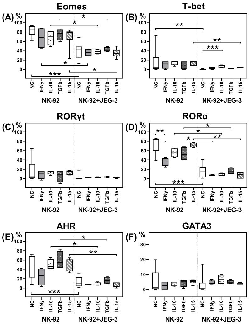
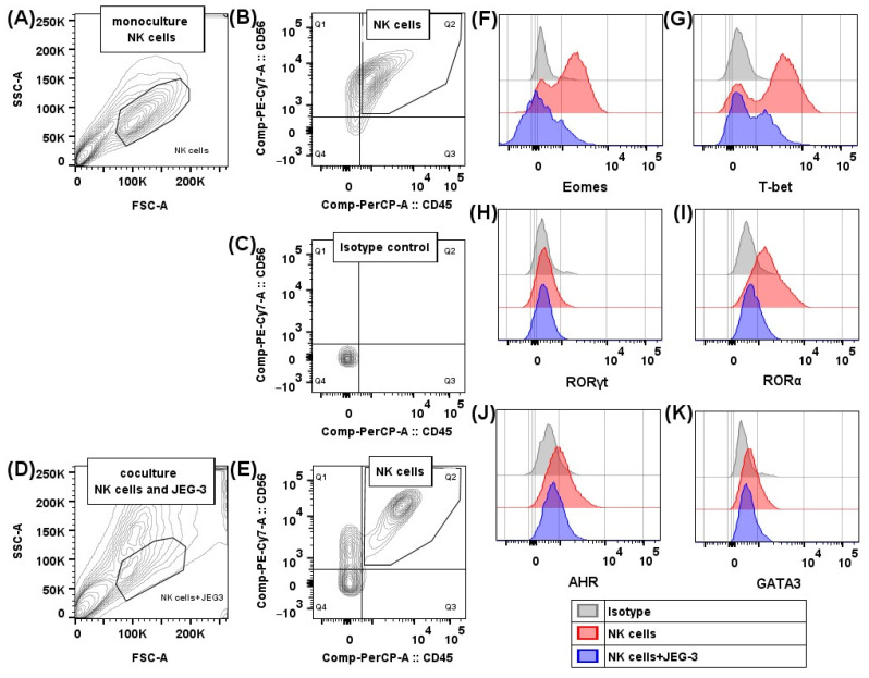
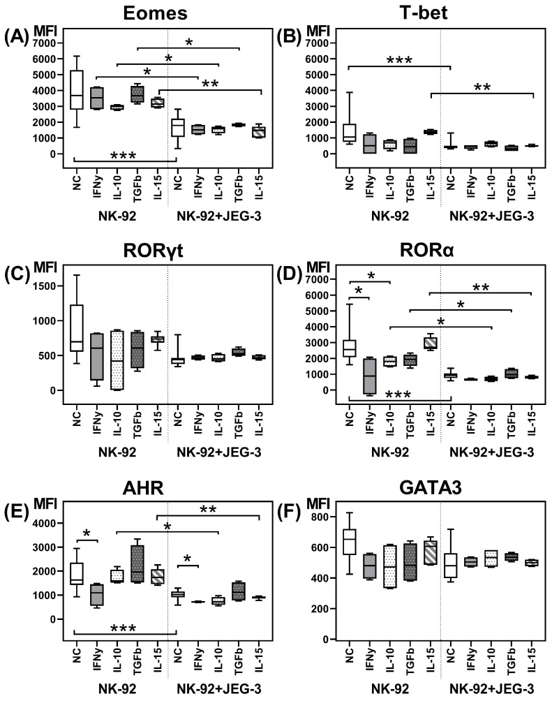
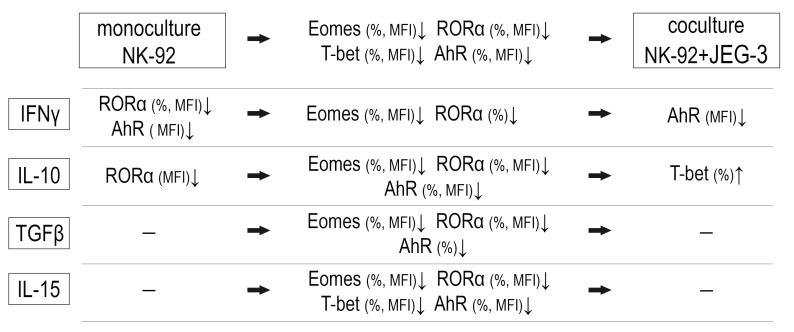
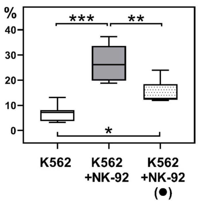
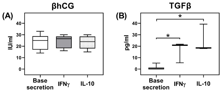
The uterine decidua contains NK cells differing in their characteristics from classical NK cells, as well as other populations of innate lymphoid cells (ILCs). ILC differentiation depends on the active transcription factors: ILC1 is characterized by T-bet expression, ILC2 is defined by RORα and GATA3, ILC3 expresses RORγt and AhR. We analyzed in vitro the expression of transcription factors by NK cells in the presence of trophoblast cells and cytokines and changes in NK cell cytotoxic activity. We used NK-92 and JEG-3 cell lines, which we cocultured in the presence of IFNγ, IL-10, IL-15, and TGFβ. Then, cells were treated with antibodies to AhR, Eomes, GATA-3, RORα, RORγt, and T-bet and were analyzed. We determined NK cell cytotoxicity towards K562 cells. To characterize the functional state of trophoblast cells, we estimated their secretion of TGFβ and βhCG. We showed that in the presence of trophoblasts, the expression of the classical NK cell transcription factors-Eomes, T-bet, as well as RORα, regulating ILC2 differentiation, and AhR, participating in NCR+ ILC3 formation-decreased in NK cells. RORγt expression typical for NCR- ILC3 remained unchanged. IFNγ inhibited AhR expression. IL-10 stimulated an increase in the number of T-bet+ ILC1-like cells. Both IL-10 and IFNγ suppressed RORα expression by NK cells and stimulated TGFβ secretion by trophoblasts. After coculture with trophoblast cells, NK cells reduced their cytotoxicity. These results indicated trophoblast cell influence on the acquisition of ILC1 and ILC3 characteristics by NK cells.
子宫蜕膜含有特性不同于经典自然杀伤(NK)细胞的NK细胞,以及其他固有淋巴细胞(ILC)群体。ILC的分化取决于活性转录因子:ILC1的特征是表达T-bet,ILC2由RORα和GATA3定义,ILC3表达RORγt和芳烃受体(AhR)。我们在体外分析了滋养层细胞和细胞因子存在时NK细胞转录因子的表达以及NK细胞细胞毒性活性的变化。我们使用了NK-92和JEG-3细胞系,将它们在干扰素γ(IFNγ)、白细胞介素10(IL-10)、白细胞介素15(IL-15)和转化生长因子β(TGFβ)存在的情况下进行共培养。然后,用针对AhR、Eomes、GATA-3、RORα、RORγt和T-bet的抗体处理细胞并进行分析。我们测定了NK细胞对K562细胞的细胞毒性。为了表征滋养层细胞的功能状态,我们评估了它们TGFβ和β人绒毛膜促性腺激素(βhCG)的分泌。我们发现,在存在滋养层细胞的情况下,经典NK细胞转录因子——Eomes、T-bet以及调节ILC2分化的RORα和参与NCR+ ILC3形成的AhR在NK细胞中的表达降低。NCR- ILC3典型的RORγt表达保持不变。IFNγ抑制AhR表达。IL-10刺激T-bet+ ILC1样细胞数量增加。IL-10和IFNγ均抑制NK细胞的RORα表达并刺激滋养层细胞分泌TGFβ。与滋养层细胞共培养后,NK细胞降低了它们的细胞毒性。这些结果表明滋养层细胞对NK细胞获得ILC1和ILC3特征有影响。