Lee Sang Eun, Joo Jeong Ho, Hwang Hee Sang, Chen Shang-Fu, Evans Douglas, Lee Kyoung Yul, Kim Kyung-Hee, Hyun Junho, Kim Min-Seok, Jung Sung-Ho, Kim Jae-Joong, Lee Jeong Seok, Torkamani Ali
Department of Cardiology, Asan Medical Center, University of Ulsan College of Medicine, Seoul, Korea.
Scripps Research Translational Institute, 3344 North Torrey Pines Court, La Jolla, CA 92037, USA.
Eur Heart J. 2025 Aug 14;46(31):3098-3114. doi: 10.1093/eurheartj/ehaf272.
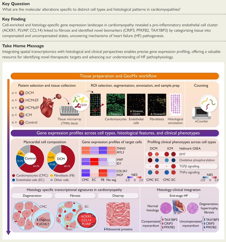
Heart failure (HF) remains a significant clinical challenge due to its diverse aetiologies and complex pathophysiology. The molecular alterations specific to distinct cell types and histological patterns during HF progression are still poorly characterized. This study aimed to explore cell-type- and histology-specific gene expression profiles in cardiomyopathies.
Ninety tissue cores from 44 participants, encompassing various forms of cardiomyopathy and control samples with diverse histological features, were analysed using the GeoMx Whole Human Transcriptome Atlas. Data on cell types, clinical information, and histological features were integrated to examine gene expression profiles in cardiomyopathy.
The study characterized the cellular composition of ventricular myocardium and validated the GeoMx platform's efficiency in compartmentalizing specific cell types, demonstrating high accuracy for cardiomyocytes but limitations for endothelial cells and fibroblasts. Differentially expressed genes, including UCHL1 from cardiomyocytes, were associated with degeneration, while CCL14, ACKR1, and PLVAP from endothelial cells were linked to fibrosis. Multiplex immunohistochemistry and integrative analysis of prior sc/snRNA-seq data identified a PLVAP, ACKR1, and CCL14-positive pro-inflammatory endothelial cell subtype linked to fibrosis in HF. Downregulation of ribosomal proteins in cardiomyocytes was associated with myocyte disarray in hypertrophic cardiomyopathy. Additionally, pronounced inflammatory responses were observed in end-stage HF. Combined histological and clinical analysis identified CRIP3, PFKFB2, and TAX1BP3 as novel contributors to HF pathogenesis.
These findings highlight the critical role of cell-enriched and histology-specific transcriptome mapping in understanding the complex pathophysiological landscape of failing hearts, offering molecular insights and potential therapeutic targets for future interventions.
心力衰竭(HF)因其病因多样和病理生理复杂,仍然是一项重大的临床挑战。HF进展过程中不同细胞类型和组织学模式特有的分子改变仍未得到充分表征。本研究旨在探索心肌病中细胞类型和组织学特异性的基因表达谱。
使用GeoMx全人类转录组图谱分析了来自44名参与者的90个组织芯,包括各种形式的心肌病和具有不同组织学特征的对照样本。整合细胞类型、临床信息和组织学特征的数据,以检查心肌病中的基因表达谱。
该研究表征了心室心肌的细胞组成,并验证了GeoMx平台在区分特定细胞类型方面的效率,证明对心肌细胞具有高准确性,但对内皮细胞和成纤维细胞存在局限性。差异表达基因,包括来自心肌细胞的UCHL1,与变性相关,而来自内皮细胞的CCL14、ACKR1和PLVAP与纤维化相关。多重免疫组织化学和先前sc/snRNA-seq数据的综合分析确定了一种与HF纤维化相关的PLVAP、ACKR1和CCL14阳性促炎内皮细胞亚型。心肌细胞中核糖体蛋白的下调与肥厚型心肌病中的心肌细胞紊乱有关。此外,在终末期HF中观察到明显的炎症反应。组织学和临床联合分析确定CRIP3、PFKFB2和TAX1BP3是HF发病机制的新贡献者。
这些发现突出了细胞富集和组织学特异性转录组图谱在理解衰竭心脏复杂病理生理格局中的关键作用,为未来干预提供了分子见解和潜在治疗靶点。