Department of Commercializing Organoid Technology, NEXEL Co., Ltd., Seoul, 07802, Korea.
Centre for Research, Hudson Institute of Medical Research, Monash University, Clayton, VIC, 3168, Australia.
Cell Death Dis. 2024 May 1;15(5):308. doi: 10.1038/s41419-024-06703-9.
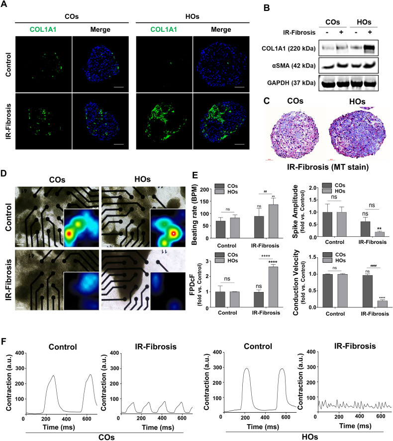
Heart disease involves irreversible myocardial injury that leads to high morbidity and mortality rates. Numerous cell-based cardiac in vitro models have been proposed as complementary approaches to non-clinical animal research. However, most of these approaches struggle to accurately replicate adult human heart conditions, such as myocardial infarction and ventricular remodeling pathology. The intricate interplay between various cell types within the adult heart, including cardiomyocytes, fibroblasts, and endothelial cells, contributes to the complexity of most heart diseases. Consequently, the mechanisms behind heart disease induction cannot be attributed to a single-cell type. Thus, the use of multi-cellular models becomes essential for creating clinically relevant in vitro cell models. This study focuses on generating self-organizing heart organoids (HOs) using human-induced pluripotent stem cells (hiPSCs). These organoids consist of cardiomyocytes, fibroblasts, and endothelial cells, mimicking the cellular composition of the human heart. The multi-cellular composition of HOs was confirmed through various techniques, including immunohistochemistry, flow cytometry, q-PCR, and single-cell RNA sequencing. Subsequently, HOs were subjected to hypoxia-induced ischemia and ischemia-reperfusion (IR) injuries within controlled culture conditions. The resulting phenotypes resembled those of acute myocardial infarction (AMI), characterized by cardiac cell death, biomarker secretion, functional deficits, alterations in calcium ion handling, and changes in beating properties. Additionally, the HOs subjected to IR efficiently exhibited cardiac fibrosis, displaying collagen deposition, disrupted calcium ion handling, and electrophysiological anomalies that emulate heart disease. These findings hold significant implications for the advancement of in vivo-like 3D heart and disease modeling. These disease models present a promising alternative to animal experimentation for studying cardiac diseases, and they also serve as a platform for drug screening to identify potential therapeutic targets.
心脏病涉及不可逆转的心肌损伤,导致高发病率和死亡率。已经提出了许多基于细胞的心脏体外模型作为非临床动物研究的补充方法。然而,这些方法中的大多数都难以准确复制成人心脏的条件,如心肌梗死和心室重构病理学。成年心脏内各种细胞类型之间的复杂相互作用,包括心肌细胞、成纤维细胞和内皮细胞,导致大多数心脏病的复杂性。因此,心脏病诱导的机制不能归因于单一细胞类型。因此,使用多细胞模型对于创建临床相关的体外细胞模型至关重要。本研究重点使用人诱导多能干细胞 (hiPSC) 生成自组织心脏类器官 (HO)。这些类器官包含心肌细胞、成纤维细胞和内皮细胞,模拟人类心脏的细胞组成。通过各种技术,包括免疫组织化学、流式细胞术、q-PCR 和单细胞 RNA 测序,证实了 HOs 的多细胞组成。随后,在受控培养条件下,将 HOs 暴露于缺氧诱导的缺血和缺血再灌注 (IR) 损伤下。由此产生的表型类似于急性心肌梗死 (AMI),其特征是心脏细胞死亡、生物标志物分泌、功能缺陷、钙离子处理改变和搏动特性改变。此外,IR 处理的 HOs 有效地表现出心脏纤维化,显示胶原蛋白沉积、钙离子处理中断以及模拟心脏病的电生理异常。这些发现对推进类似于体内的 3D 心脏和疾病建模具有重要意义。这些疾病模型为研究心脏疾病提供了一种有前途的动物实验替代方法,并且它们也是筛选药物以识别潜在治疗靶点的平台。