Zhang Xiaolei, Chen Xiaoyan, Fu Yao, Zhou Han, Lin Yan
Department of Radiology, Second Affiliated Hospital of Shantou University Medical College, No. 69, Dongxiabei Road, Jinping District, Shantou, Guangdong Province, 515041, China.
BMC Med Imaging. 2025 May 13;25(1):159. doi: 10.1186/s12880-025-01698-x.
This study aimed to visually analyze the heterogeneity of vascularity and cellularity across different sub-regions of breast cancer using habitat imaging (HI) to predict human epidermal growth factor receptor 2 (HER2) expression and evaluate the effectiveness of neoadjuvant therapy (NAT) in breast cancer patients.
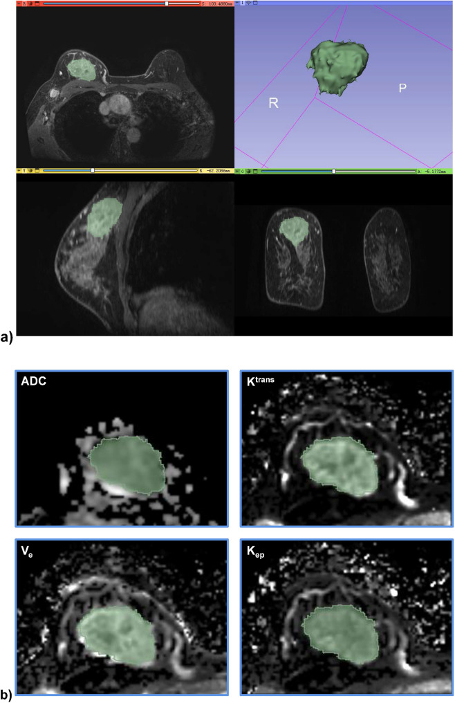
A retrospective analysis was conducted on 76 patients diagnosed with breast cancer. Diffusion-weighted imaging (DWI) and dynamic contrast-enhanced MRI (DCE-MRI) sequences were utilized to acquire MR images. Apparent diffusion coefficient (ADC), K, K, and V values were measured for each sub-region, and the percentage of each sub-region relative to the total lesion was calculated. Statistical analyses, including t-tests, rank-sum tests, chi-square tests, and Spearman correlation, were performed.
Three distinct sub-regions within breast cancer lesions were identified through HI, characterized physiologically as: low vascularity-high cellularity (LV-HC), low vascularity-low cellularity (LV-LC), and high vascularity-low cellularity (HV-LC). Significant differences were observed in the proportions of these tumor sub-regions between HER2-positive and HER2-negative breast cancers. Additionally, HER2-low and HER2-zero breast cancers demonstrated statistical differences in the second sub-region (LV-LC). Furthermore, the proportion of the first sub-region (LV-HC) was negatively correlated with the efficacy of NAT in breast cancer patients.
Habitat imaging can identify distinct sub-regions within breast cancer lesions, providing a noninvasive imaging biomarker for predicting HER2 expression levels and assessing the efficacy of NAT in breast cancer patients.
本研究旨在利用栖息地成像(HI)对乳腺癌不同亚区域的血管生成和细胞构成的异质性进行可视化分析,以预测人表皮生长因子受体2(HER2)表达,并评估新辅助治疗(NAT)对乳腺癌患者的疗效。
对76例确诊为乳腺癌的患者进行回顾性分析。利用扩散加权成像(DWI)和动态对比增强磁共振成像(DCE-MRI)序列采集磁共振图像。测量每个亚区域的表观扩散系数(ADC)、K、K和V值,并计算每个亚区域相对于总病变的百分比。进行了包括t检验、秩和检验、卡方检验和Spearman相关性分析在内的统计分析。
通过HI在乳腺癌病灶内确定了三个不同的亚区域,其生理特征为:低血管生成-高细胞构成(LV-HC)、低血管生成-低细胞构成(LV-LC)和高血管生成-低细胞构成(HV-LC)。HER2阳性和HER2阴性乳腺癌在这些肿瘤亚区域的比例上存在显著差异。此外,HER2低表达和HER2零表达乳腺癌在第二个亚区域(LV-LC)表现出统计学差异。此外,第一个亚区域(LV-HC)的比例与乳腺癌患者NAT的疗效呈负相关。
栖息地成像可以识别乳腺癌病灶内不同的亚区域,为预测HER2表达水平和评估NAT对乳腺癌患者的疗效提供一种非侵入性成像生物标志物。