Wu Qiulin, Yang Yan, Lin Shixun, Geller David A, Yan Yihe
Department of General Surgery, The Second Affiliated Hospital of Guangxi Medical University, Nanning, Guangxi, China.
Thomas E. Starzl Transplantation Institute, Department of Surgery, University of Pittsburgh Medical Center, Pittsburgh, PA, United States.
Front Immunol. 2025 Apr 30;16:1569915. doi: 10.3389/fimmu.2025.1569915. eCollection 2025.
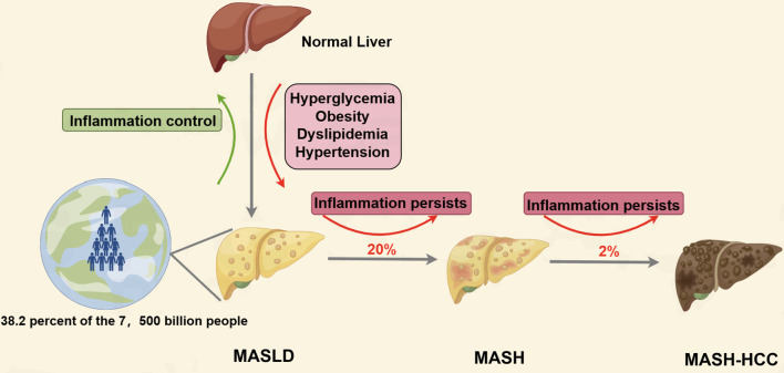
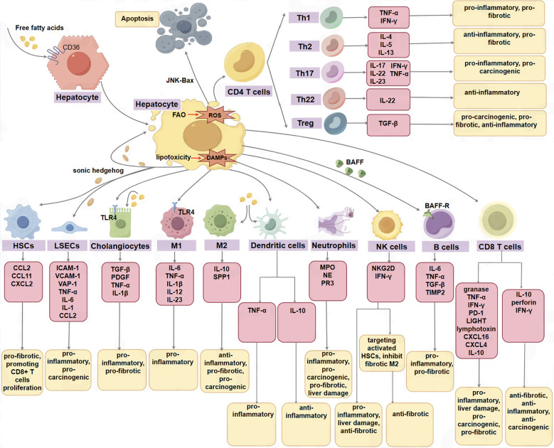
Metabolic dysfunction-associated steatotic liver disease (MASLD) is a series of obesity-related metabolic liver diseases, ranging from relatively benign hepatic steatosis to metabolic-associated steatohepatitis (MASH). With the changes in lifestyle, its incidence and prevalence have risen to epidemic proportions globally. In recent years, an increasing amount of evidence has indicated that the hepatic microenvironment is involved in the pathophysiological processes of MASH-induced liver fibrosis and the formation of hepatocellular carcinoma (HCC). The hepatic microenvironment is composed of various parenchymal and non-parenchymal cells, which communicate with each other through various factors. In this review, we focus on the changes in hepatocytes, cholangiocytes, liver sinusoidal endothelial cells (LSECs), hepatic stellate cells (HSCs), Kupffer cells (KC), dendritic cells (DC), neutrophils, monocytes, T and B lymphocytes, natural killer cells (NK), natural killer T cells (NKT), mucosal-associated invariant T cells (MAIT), γδT cells, and gut microbiota during the progression of MASLD. Furthermore, we discuss promising therapeutic strategies targeting the microenvironment of MASLD-MASH-HCC.
代谢功能障碍相关脂肪性肝病(MASLD)是一系列与肥胖相关的代谢性肝病,范围从相对良性的肝脂肪变性到代谢相关脂肪性肝炎(MASH)。随着生活方式的改变,其发病率和患病率在全球范围内已上升至流行程度。近年来,越来越多的证据表明,肝脏微环境参与了MASH诱导的肝纤维化和肝细胞癌(HCC)形成的病理生理过程。肝脏微环境由各种实质细胞和非实质细胞组成,它们通过各种因子相互交流。在本综述中,我们重点关注MASLD进展过程中肝细胞、胆管细胞、肝窦内皮细胞(LSEC)、肝星状细胞(HSC)、库普弗细胞(KC)、树突状细胞(DC)、中性粒细胞、单核细胞、T和B淋巴细胞、自然杀伤细胞(NK)、自然杀伤T细胞(NKT)、黏膜相关恒定T细胞(MAIT)、γδT细胞和肠道微生物群的变化。此外,我们还讨论了针对MASLD-MASH-HCC微环境的有前景的治疗策略。