Zhou Yuan, Zhou Liang, Wang Meng, Xu Lin, Li Tingting, Wang Chen
Department of Nephrology, Shuguang Hospital Affiliated to Shanghai University of Traditional Chinese Medicine, Shanghai, China.
Key Laboratory of Liver and Kidney Diseases, Ministry of Education, Shanghai University of Traditional Chinese Medicine, Shanghai, China.
Ren Fail. 2025 Dec;47(1):2502875. doi: 10.1080/0886022X.2025.2502875. Epub 2025 May 19.
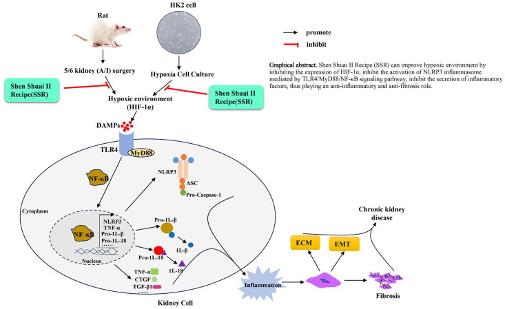
To investigate the anti-fibrotic mechanisms of Shen Shuai II Recipe (SSR) in chronic kidney disease (CKD), focusing on its modulation of hypoxia-associated inflammatory pathways and the TLR4/MyD88/NF-κB/NLRP3 axis.
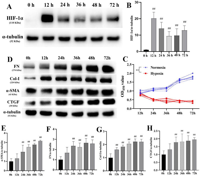
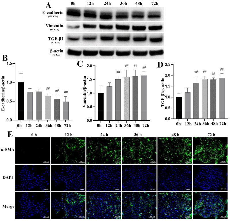
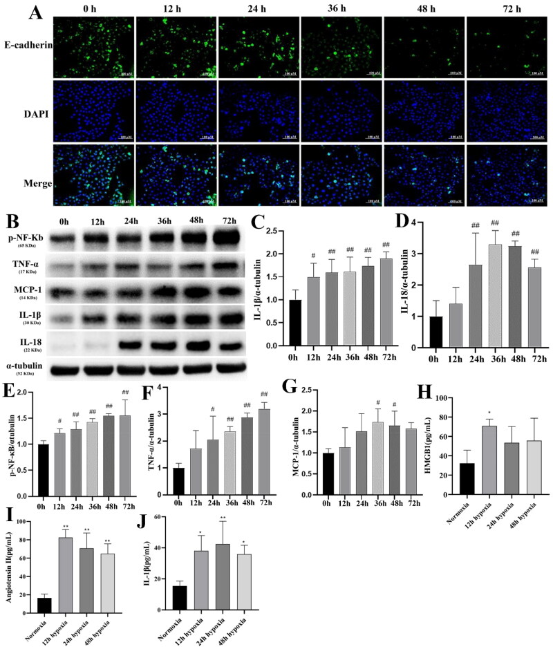
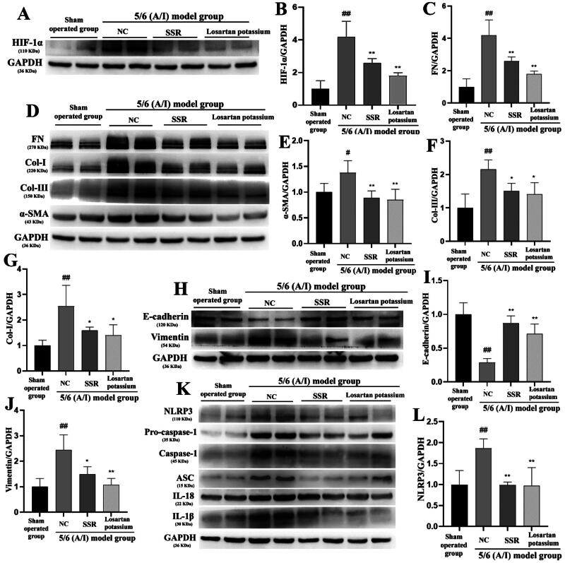
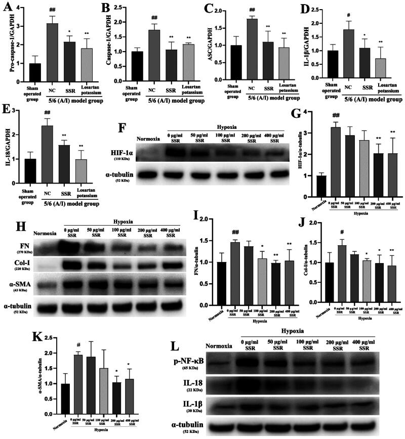
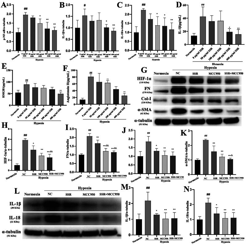
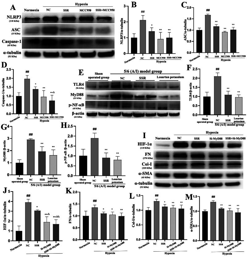
A 5/6 nephrectomy-induced chronic renal failure (CRF) rat model and hypoxia-exposed human renal tubular epithelial (HK-2) cells were utilized. In vivo, renal function was assessed via serum creatinine, urea nitrogen, and creatinine clearance measurements, alongside histopathological evaluation of renal fibrosis and inflammation. In vitro, hypoxia-treated HK-2 cells were analyzed for fibrotic markers (fibronectin, collagen I, α-smooth muscle actin) and pro-inflammatory cytokines (IL-1β, IL-18). Molecular mechanisms were probed through protein expression analysis of HIF-1α and the TLR4/MyD88/NF-κB pathway, with NLRP3 inflammasome activity evaluated.
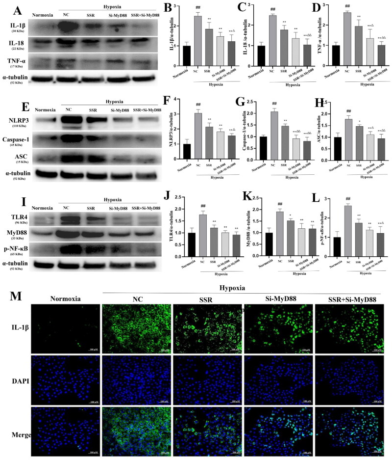
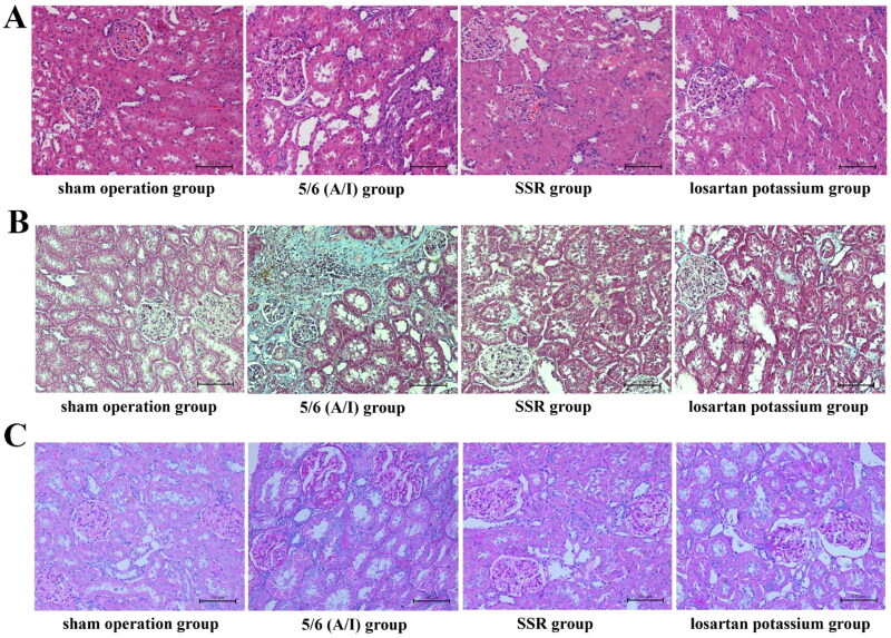
SSR treatment significantly improved renal function in CRF rats, reducing serum creatinine (Scr) and urea nitrogen (BUN) while enhancing creatinine clearance. Histopathology revealed preserved renal architecture with attenuated fibrosis and inflammatory infiltration. In hypoxic HK-2 cells, SSR downregulated fibrotic markers and suppressed IL-1β and IL-18 levels. Mechanistically, SSR reduced HIF-1α expression, inhibited TLR4/MyD88/NF-κB signaling, and suppressed NLRP3 inflammasome activation in both models.
SSR alleviates renal fibrosis and CKD progression by mitigating hypoxia-driven inflammation and blocking the TLR4/MyD88/NF-κB/NLRP3 pathway.
研究肾衰Ⅱ号方(SSR)在慢性肾脏病(CKD)中的抗纤维化机制,重点关注其对缺氧相关炎症通路以及TLR4/MyD88/NF-κB/NLRP3轴的调节作用。
采用5/6肾切除诱导的慢性肾衰竭(CRF)大鼠模型和缺氧处理的人肾小管上皮(HK-2)细胞。在体内,通过测定血清肌酐、尿素氮和肌酐清除率评估肾功能,并对肾纤维化和炎症进行组织病理学评估。在体外,分析缺氧处理的HK-2细胞中的纤维化标志物(纤连蛋白、Ⅰ型胶原、α-平滑肌肌动蛋白)和促炎细胞因子(IL-1β、IL-18)。通过对HIF-1α和TLR4/MyD88/NF-κB通路的蛋白表达分析探究分子机制,并评估NLRP3炎性小体活性。
SSR治疗显著改善了CRF大鼠的肾功能,降低了血清肌酐(Scr)和尿素氮(BUN),同时提高了肌酐清除率。组织病理学显示肾脏结构保存,纤维化和炎症浸润减轻。在缺氧的HK-2细胞中,SSR下调了纤维化标志物并抑制了IL-1β和IL-18水平。机制上,SSR在两种模型中均降低了HIF-1α表达,抑制了TLR4/MyD88/NF-κB信号传导,并抑制了NLRP3炎性小体激活。
SSR通过减轻缺氧驱动的炎症并阻断TLR4/MyD88/NF-κB/NLRP3通路,减轻肾纤维化和CKD进展。