Xu Tian-Ye, Feng Yan-Hong, Sun Zhong-Ru, He Liang, Chen Jin-Hua, Tian Wei-Zhong, Zhang Hong-Xia, Zhu Meng, Xia Jian-Guo
Graduate School of Dalian Medical University, Dalian, China.
Department of Imaging, Taizhou People's Hospital, Taizhou, China.
Quant Imaging Med Surg. 2025 May 1;15(5):3982-3992. doi: 10.21037/qims-24-1440. Epub 2025 Apr 8.
Cognitive decline may occur in patients with diabetic retinopathy (DR), yet the mechanism underlying the relationship between cognitive decline and DR remains unclear. This study applied an automated fiber-tract quantification (AFQ) technique based on diffusion tensor imaging (DTI) to identify alterations in specific segments of brain white matter fiber tracts in patients with DR, and analyze their correlation with cognitive test scores and clinical biochemical indicators.
A total of 19 patients with DR and 20 age-, sex-, and education-matched healthy controls (HCs) were included. Clinical and imaging data were prospectively collected. The AFQ technique was applied to track the whole brain white matter fiber tracts of each participant, and each fiber tract was segmented into 100 equidistant nodes. The fractional anisotropy (FA), mean diffusion (MD), axial diffusion (AD), and radial diffusion in 100 nodes of each fiber tract were calculated and compared between the two groups. Partial correlation analysis was performed to analyze the correlation between altered DTI metrics in segments of the fiber tracts and cognitive test scores, as well as clinical biochemical indicators in patients with DR.
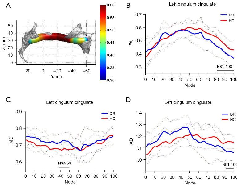
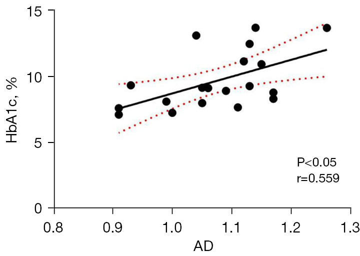
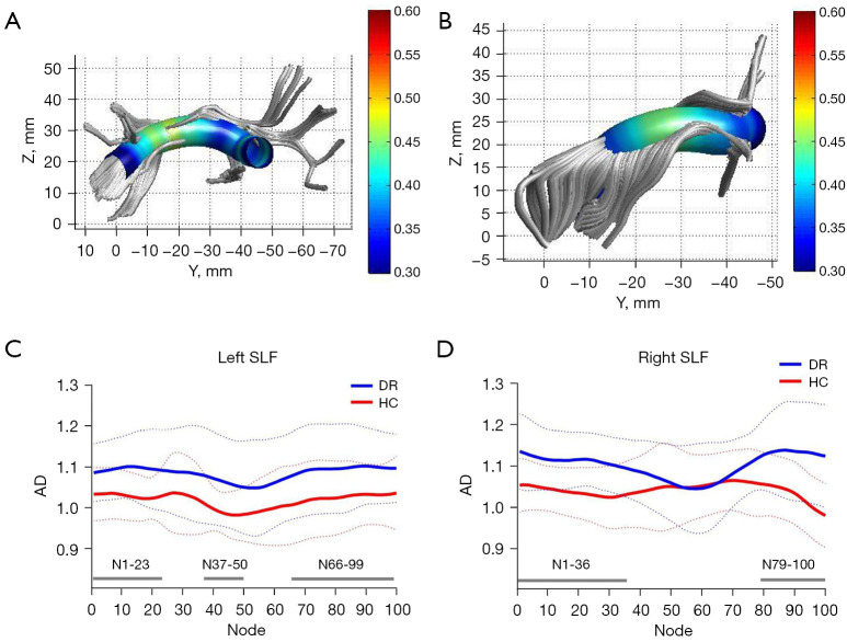
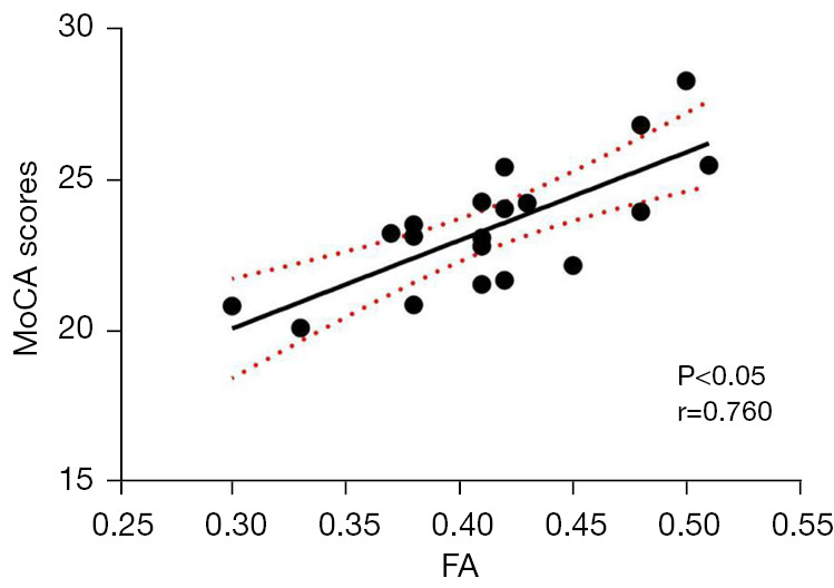
Compared with the HC group, the DR group showed significantly reduced FA values in nodes 81-100, increased MD values in nodes 39-50, and reduced AD values in nodes 91-100 of the left cingulum cingulate (CGC) [P<0.05, false discovery rate (FDR) corrected], they also showed increased AD values in the left superior longitudinal fasciculus (SLF; nodes 1-23, 37-50, and 66-99), and the right SLF (nodes 1-36 and 79-100) (P<0.05, FDR corrected). Correlation analysis revealed a positive correlation between the FA values in nodes 82-98 of the left CGC and Montreal Cognitive Assessment scores (MoCA scores, r=0.760, P<0.05/P=0.021), and a positive correlation between the AD values in nodes 37-41 in the left SLF and glycated hemoglobin A1c (HbA1c) levels (r=0.559, P<0.05/P=0.039).
Our findings demonstrated alterations in the white matter fiber tracts at the point-wise level in patients with DR using AFQ analysis. These alterations may be associated with cognitive impairment in DR. The AFQ technique can accurately detect the damage to the integrity of the brain white matter fiber tracts in patients with DR, and have high clinical application value in the diagnosis and evaluation of DR, which can deepen our understanding of brain white matter microstructural abnormalities in patients with DR.
糖尿病视网膜病变(DR)患者可能会出现认知功能下降,但其与DR之间关系的潜在机制尚不清楚。本研究应用基于扩散张量成像(DTI)的自动纤维束定量(AFQ)技术,以识别DR患者脑白质纤维束特定节段的改变,并分析其与认知测试分数和临床生化指标的相关性。
共纳入19例DR患者和20例年龄、性别和教育程度相匹配的健康对照者(HCs)。前瞻性收集临床和影像数据。应用AFQ技术追踪每位参与者的全脑白质纤维束,并将每条纤维束分成100个等距节点。计算并比较两组每条纤维束100个节点处的分数各向异性(FA)、平均扩散率(MD)、轴向扩散率(AD)和径向扩散率。进行偏相关分析,以分析纤维束节段中DTI指标改变与DR患者认知测试分数以及临床生化指标之间的相关性。
与HC组相比,DR组左侧扣带束(CGC)81-100节点处FA值显著降低,39-50节点处MD值升高,91-100节点处AD值降低[P<0.05,错误发现率(FDR)校正],他们还显示左侧上纵束(SLF;1-23、37-50和66-99节点)以及右侧SLF(1-36和79-100节点)的AD值升高(P<0.05,FDR校正)。相关性分析显示,左侧CGC的82-98节点处FA值与蒙特利尔认知评估分数(MoCA分数,r=0.760,P<0.05/P=0.021)呈正相关,左侧SLF的37-41节点处AD值与糖化血红蛋白A1c(HbA1c)水平呈正相关(r=0.559,P<0.05/P=0.039)。
我们的研究结果表明,使用AFQ分析可发现DR患者白质纤维束在逐点水平上的改变。这些改变可能与DR患者的认知障碍有关。AFQ技术能够准确检测DR患者脑白质纤维束完整性的损伤,在DR的诊断和评估中具有较高的临床应用价值,可加深我们对DR患者脑白质微观结构异常的理解。