Mao Ya-Qing, Sun Rui, Liu Shuo, Zhang Wen-Bo, Yu Yao, Jia Ling-Fei, Yu Guang-Yan, Peng Xin
Department of Oral and Maxillofacial Surgery, Peking University School and Hospital of Stomatology, Beijing, P.R. China.
National Center for Stomatology, National Clinical Research Center for Oral Diseases, National Engineering Research Center of Oral Biomaterials and Digital Medical Devices, Beijing, P.R. China.
Clin Med Insights Oncol. 2025 May 16;19:11795549251335172. doi: 10.1177/11795549251335172. eCollection 2025.
The DNA methylation statuses of and show great promise as biomarkers for the detection of oral squamous cell carcinoma (OSCC). This study aims to investigate the distribution of or methylation in the exfoliated oral epithelial cells (OECs) of OSCC.
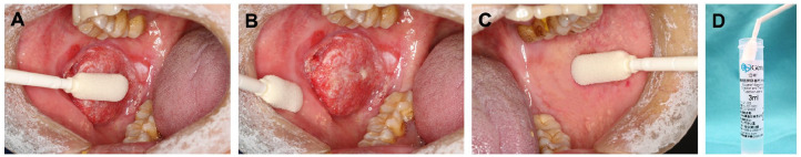
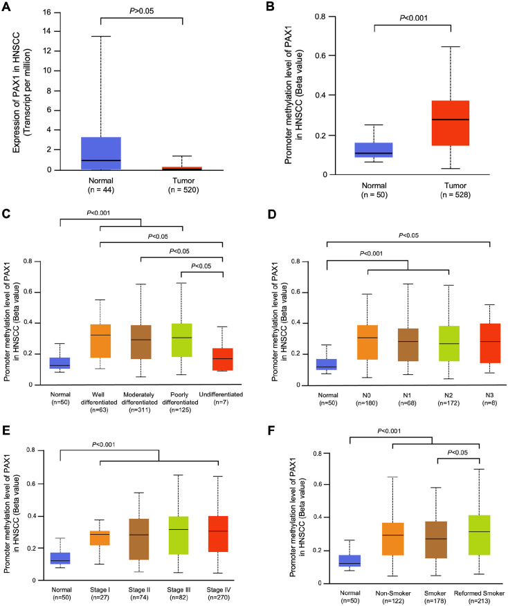
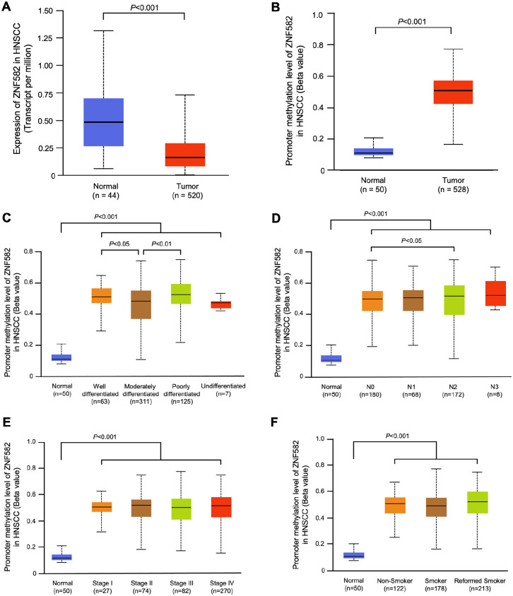
Methylation data from 528 tumors and 50 adjacent nontumor tissues were acquired from The Cancer Genome Atlas and analyzed using UALCAN database. Sixty-one OSCC cases from Peking University School and Hospital of Stomatology were included in this study and the exfoliated OECs collected by oral swabs were collected from the cancerous lesion (CL), adjacent normal (AN), and contralateral normal (CN) sites. The methylation levels of these 2 genes in different sites were evaluated.
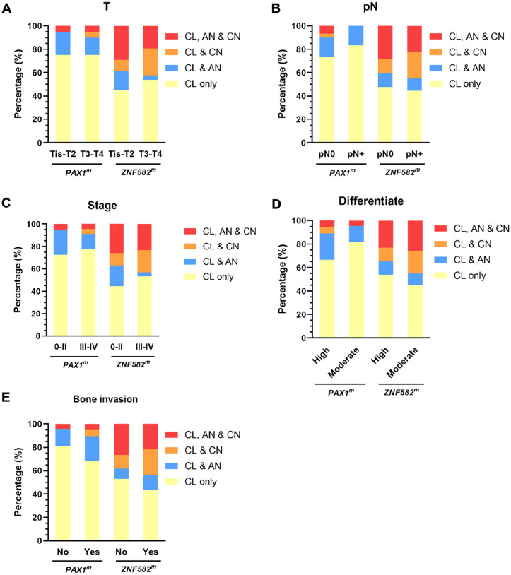
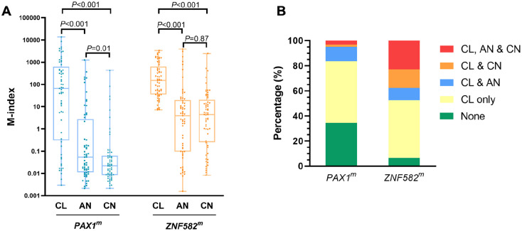
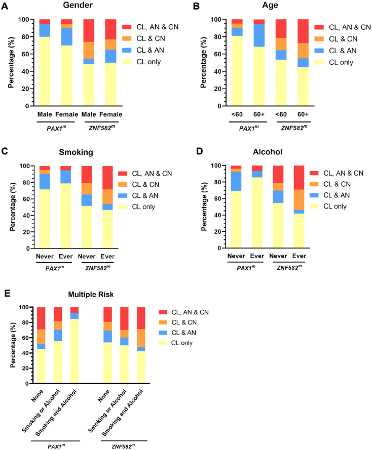
and were both hypermethylated in OSCC compared with nontumor sites but showed different methylation patterns within the oral environment. Generally, a CL-centric methylation pattern of where methylation levels decrease gradually from CL through AN to CN was observed, suggesting a field cancerization effect. methylation levels are significantly higher at lesion sites compared with normal sites, but no significant difference is observed between AN and CN. Coexistence of methylation in CL and AN or CN sites was also observed in some patients with OSCC. Furthermore, methylation was more sensitive among patients with OSCC.
DNA methylation detection of and in the exfoliated OECs is helpful for OSCC diagnosis. Hypermethylated and show different methylation patterns in the oral cavity of patients with OSCC.
[基因名称1]和[基因名称2]的DNA甲基化状态作为口腔鳞状细胞癌(OSCC)检测的生物标志物具有很大潜力。本研究旨在调查OSCC患者脱落口腔上皮细胞(OECs)中[基因名称1]或[基因名称2]甲基化的分布情况。
从癌症基因组图谱获取528个肿瘤及50个相邻非肿瘤组织的甲基化数据,并使用UALCAN数据库进行分析。本研究纳入了来自北京大学口腔医学院和口腔医院的61例OSCC病例,通过口腔拭子从癌灶(CL)、相邻正常组织(AN)和对侧正常组织(CN)部位收集脱落的OECs。评估这两个基因在不同部位的甲基化水平。
与非肿瘤部位相比,OSCC中[基因名称1]和[基因名称2]均发生高甲基化,但在口腔环境中呈现不同的甲基化模式。总体而言,观察到以CL为中心的[基因名称1]甲基化模式,即甲基化水平从CL经AN到CN逐渐降低,提示存在场癌化效应。与正常部位相比,病变部位的[基因名称2]甲基化水平显著更高,但AN和CN之间未观察到显著差异。在一些OSCC患者中还观察到CL与AN或CN部位同时存在[基因名称2]甲基化。此外,[基因名称2]甲基化在OSCC患者中更具敏感性。
检测脱落OECs中[基因名称1]和[基因名称2]的DNA甲基化有助于OSCC诊断。[基因名称1]和[基因名称2]的高甲基化在OSCC患者口腔中呈现不同的甲基化模式。