Jiang Dan, Li Ke, Sun Yining, Zhang Zicheng, Xie Shuang, Yu Xintong, Wang Ruoqi, Feng Ying, Zheng Qinxiang, Wen Yajing, Reinach Peter S, Du Yuanyuan, Zhou Meng, Chen Wei
National Clinical Research Center for Ocular Diseases, State Key Laboratory of Ophthalmology, Optometry and Visual Science, Eye Hospital, Wenzhou Medical University, Wenzhou, 325027, China.
School of Biomedical Engineering, Wenzhou Medical University, Wenzhou, 325027, China.
Genome Med. 2025 May 19;17(1):56. doi: 10.1186/s13073-025-01475-z.
The human cornea is a transparent and uniquely ordered optical-biological system. Precise coordination of its cellular mechanisms is essential to maintain its transparency and functionality. However, the spatial, cellular and molecular architecture of the human cornea and its intercellular interactions during aging have not been elucidated.
We performed single-cell RNA sequencing (scRNA-seq) and single-cell SpaTial Enhanced REsolution Omics-sequencing (scStereo-seq) analysis in corneal tissue from eight eyes of donors aged 33-88 years to elucidate the spatiotemporal cellular and molecular dynamics of human cornea aging. Immunofluorescence staining and Western blotting were performed to validate the findings.
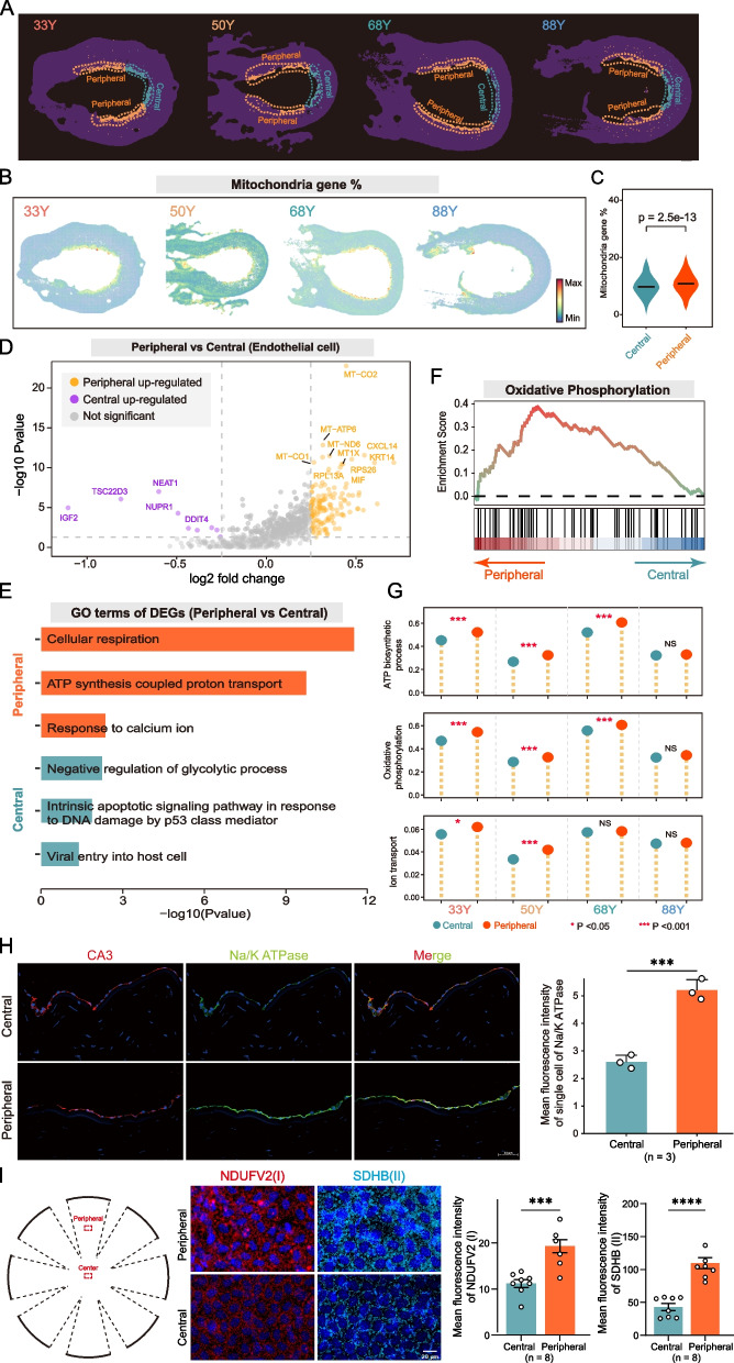
Spatiotemporal single-cell analysis revealed the complex cellular landscape, spatial organization and intercellular communication within the human cornea. The subpopulations of major cell types of the cornea were elucidated with precise spatial positions. In particular, we identified novel subpopulations, mapped the spatial positioning of limbal stem cells within the limbal niche, and delineated the interactions between major cell types. We observed that three basal cell subsets migrate centripetally from the peripheral to the central cornea with age, suggesting the "spatiotemporal centripetal pattern" as a novel paradigm for the age-related migration of corneal epithelial cells. Furthermore, we elucidated the age-related, region-specific molecular and functional characteristics of the corneal endothelium, demonstrating differential metabolic capacities and functional properties between the peripheral and central regions.
As the first comprehensive spatiotemporal atlas, our work provides a valuable resource for understanding tissue homeostasis in the human cornea and advances research on corneal pathology, transplantation, senescence and regenerative medicine in the context of corneal aging.
人类角膜是一个透明且具有独特有序结构的光学 - 生物系统。其细胞机制的精确协调对于维持其透明度和功能至关重要。然而,人类角膜的空间、细胞和分子结构及其在衰老过程中的细胞间相互作用尚未阐明。
我们对年龄在33 - 88岁的8名供体的8只眼睛的角膜组织进行了单细胞RNA测序(scRNA - seq)和单细胞空间增强分辨率组学测序(scStereo - seq)分析,以阐明人类角膜衰老的时空细胞和分子动态。进行了免疫荧光染色和蛋白质印迹分析以验证研究结果。
时空单细胞分析揭示了人类角膜内复杂的细胞景观、空间组织和细胞间通讯。明确了角膜主要细胞类型的亚群及其精确的空间位置。特别是,我们鉴定了新的亚群,绘制了角膜缘干细胞在角膜缘生态位内的空间定位,并描绘了主要细胞类型之间的相互作用。我们观察到随着年龄增长,三个基底细胞亚群从周边向中央角膜向心迁移,提示“时空向心模式”是角膜上皮细胞与年龄相关迁移的一种新范式。此外,我们阐明了角膜内皮与年龄相关的、区域特异性的分子和功能特征,证明了周边和中央区域之间不同的代谢能力和功能特性。
作为首个全面的时空图谱,我们的工作为理解人类角膜中的组织稳态提供了宝贵资源,并推动了角膜病理学、移植、衰老及角膜衰老背景下再生医学的研究。