Biosciences Institute, Faculty of Medical Sciences, Newcastle University, UK.
Newcastle Cellular Therapies Facility, Newcastle University and Newcastle Upon Tyne Hospitals NHS Foundation Trust, UK.
Ocul Surf. 2021 Jul;21:279-298. doi: 10.1016/j.jtos.2021.03.010. Epub 2021 Apr 16.
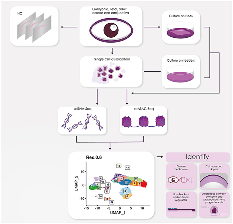
Single cell (sc) analyses of key embryonic, fetal and adult stages were performed to generate a comprehensive single cell atlas of all the corneal and adjacent conjunctival cell types from development to adulthood.
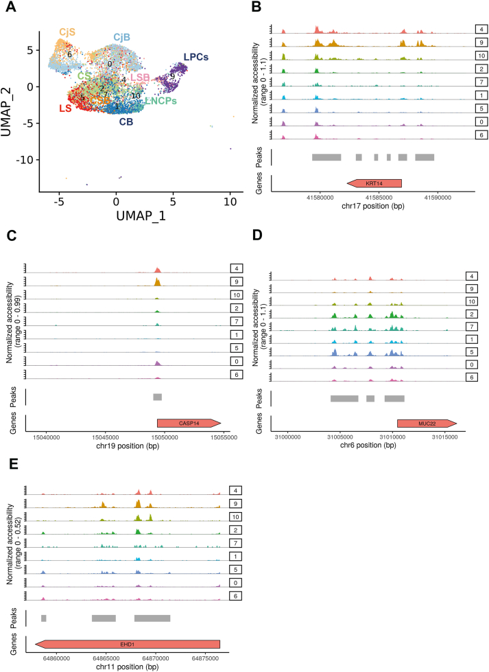
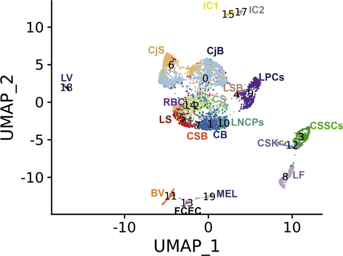
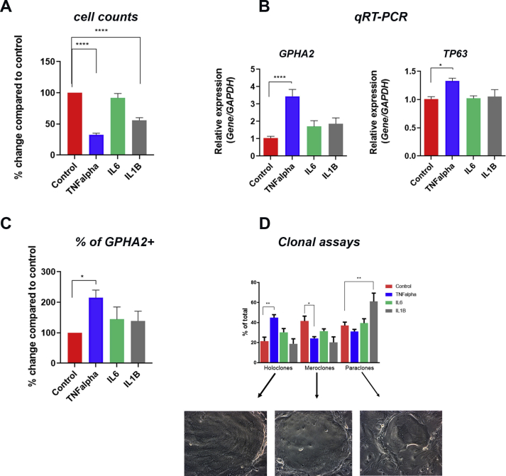
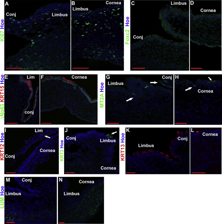
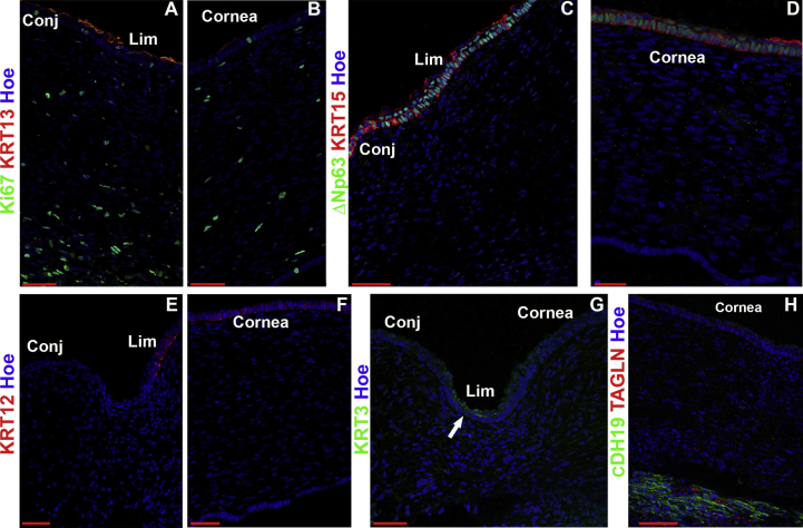
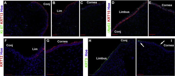
Four human adult and seventeen embryonic and fetal corneas from 10 to 21 post conception week (PCW) specimens were dissociated to single cells and subjected to scRNA- and/or ATAC-Seq using the 10x Genomics platform. These were embedded using Uniform Manifold Approximation and Projection (UMAP) and clustered using Seurat graph-based clustering. Cluster identification was performed based on marker gene expression, bioinformatic data mining and immunofluorescence (IF) analysis. RNA interference, IF, colony forming efficiency and clonal assays were performed on cultured limbal epithelial cells (LECs).
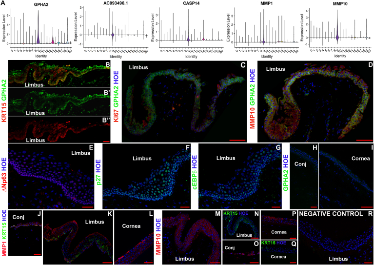
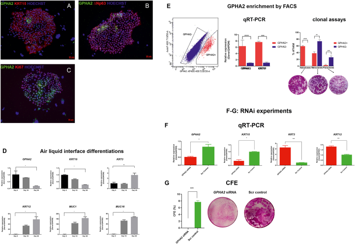
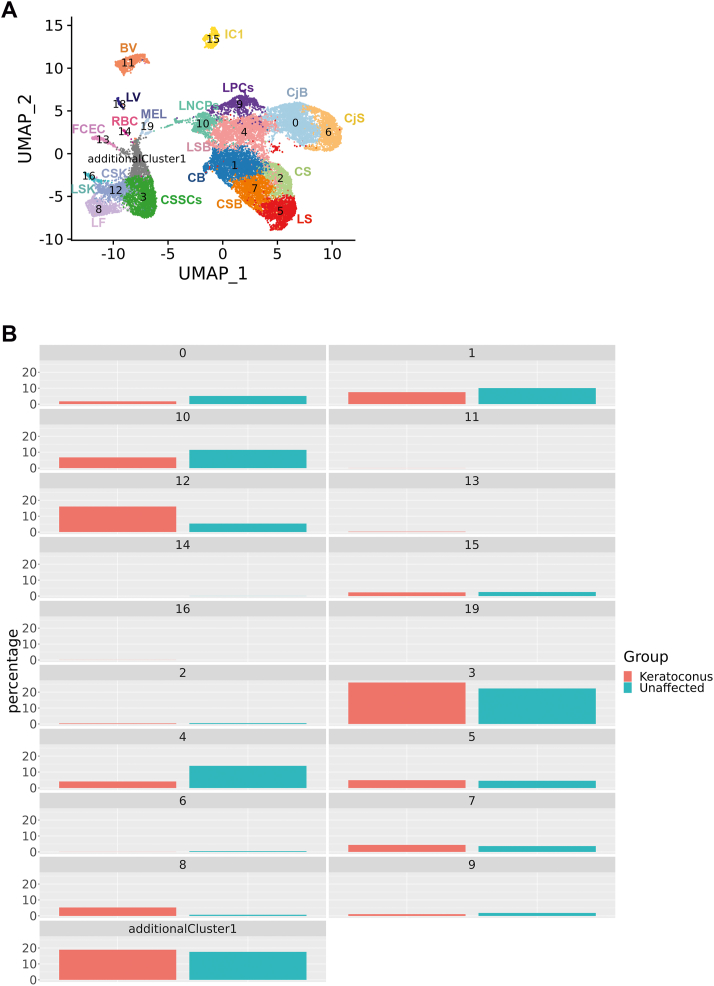
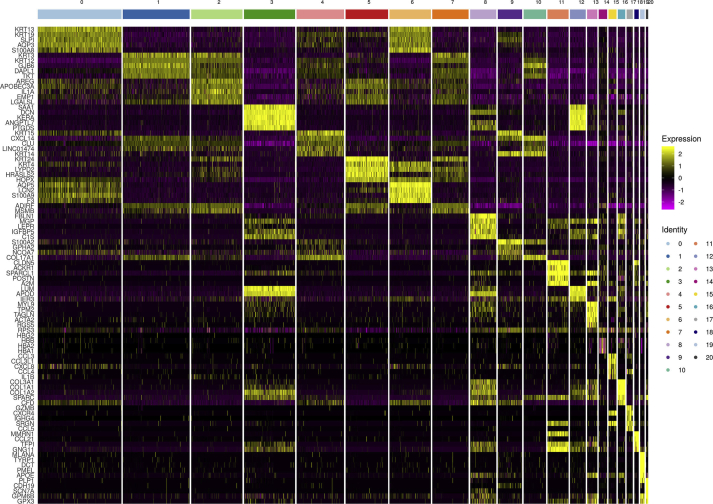
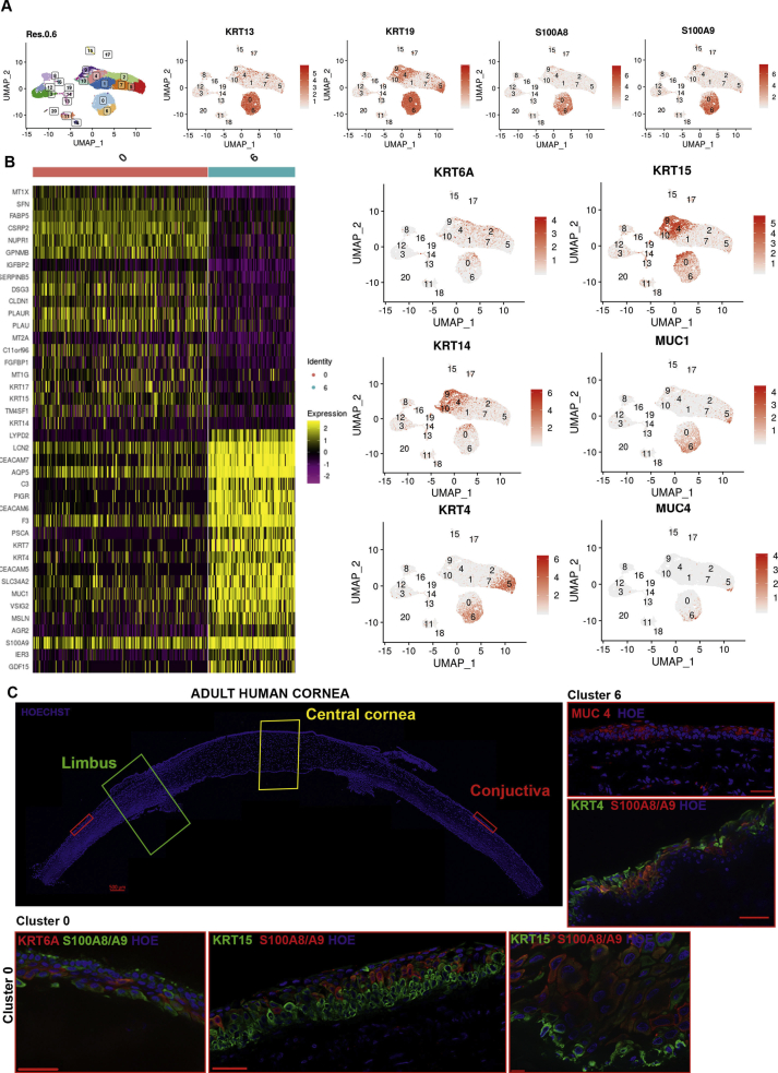
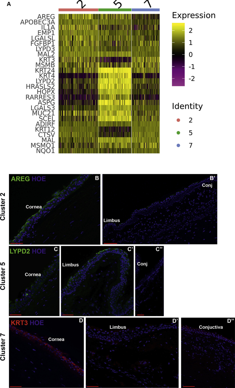
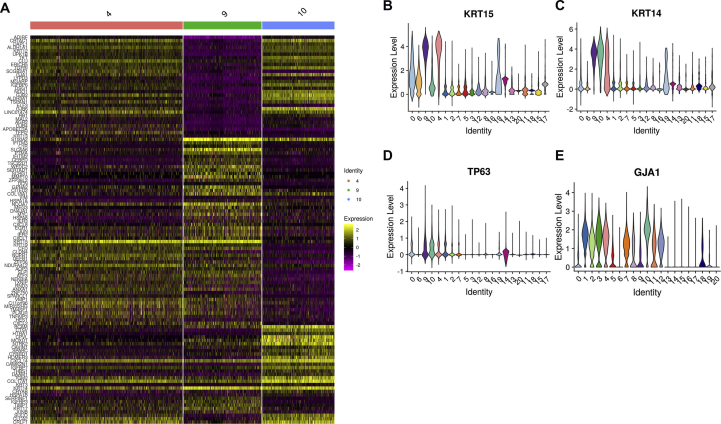
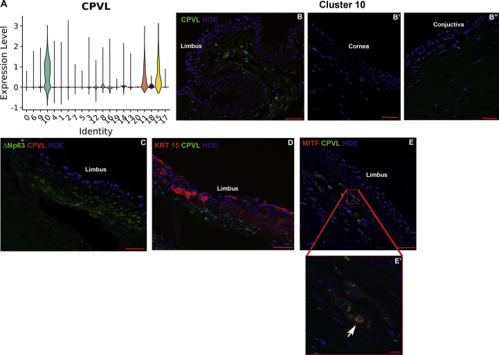
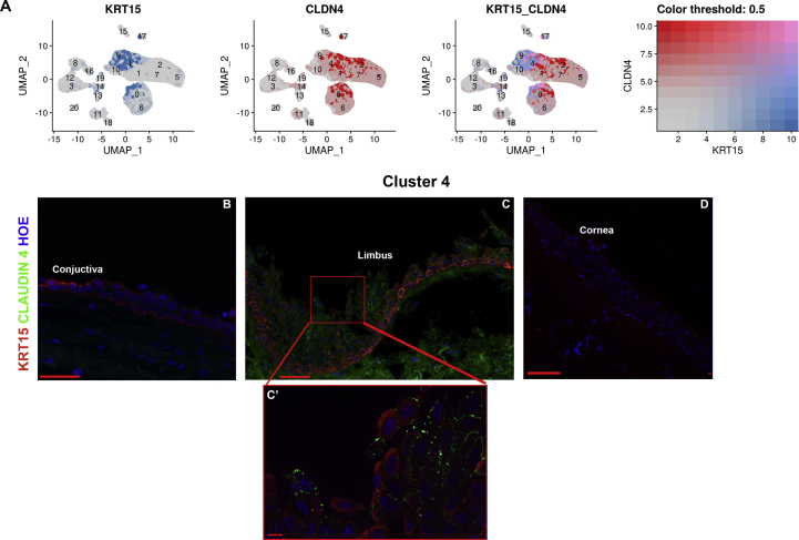
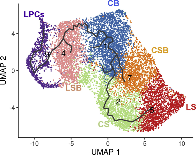
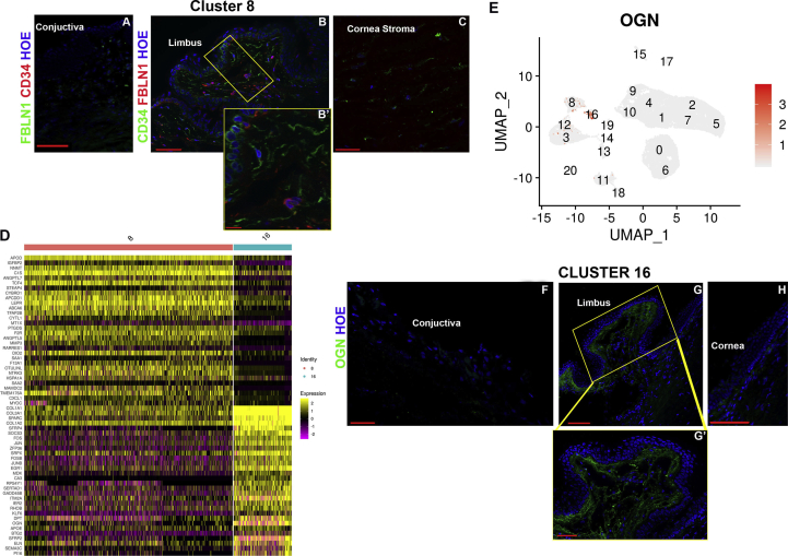
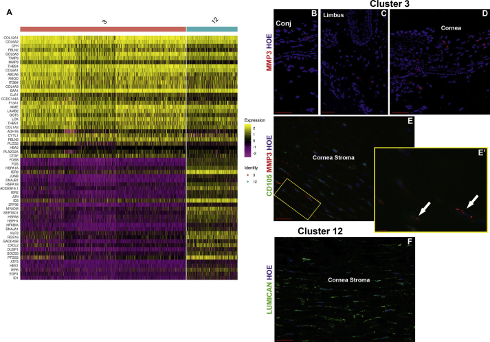
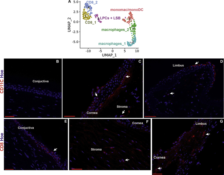
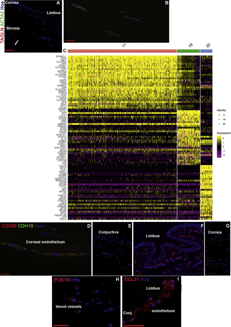
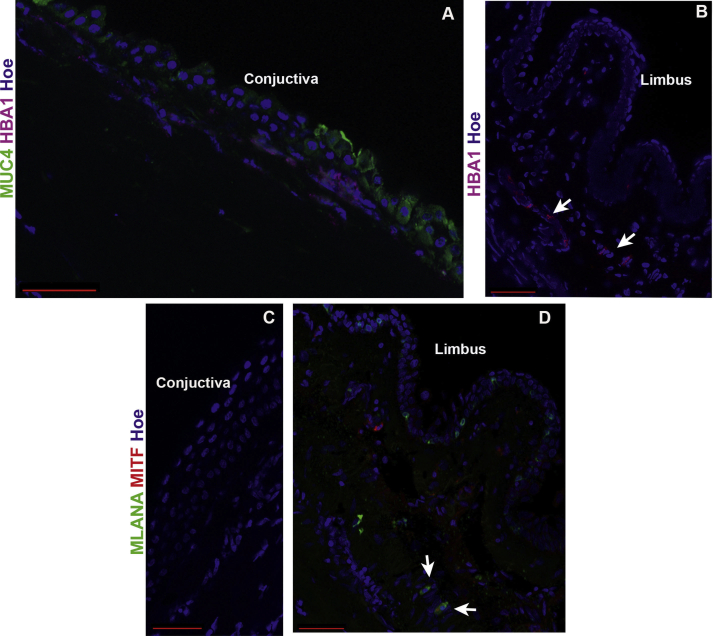
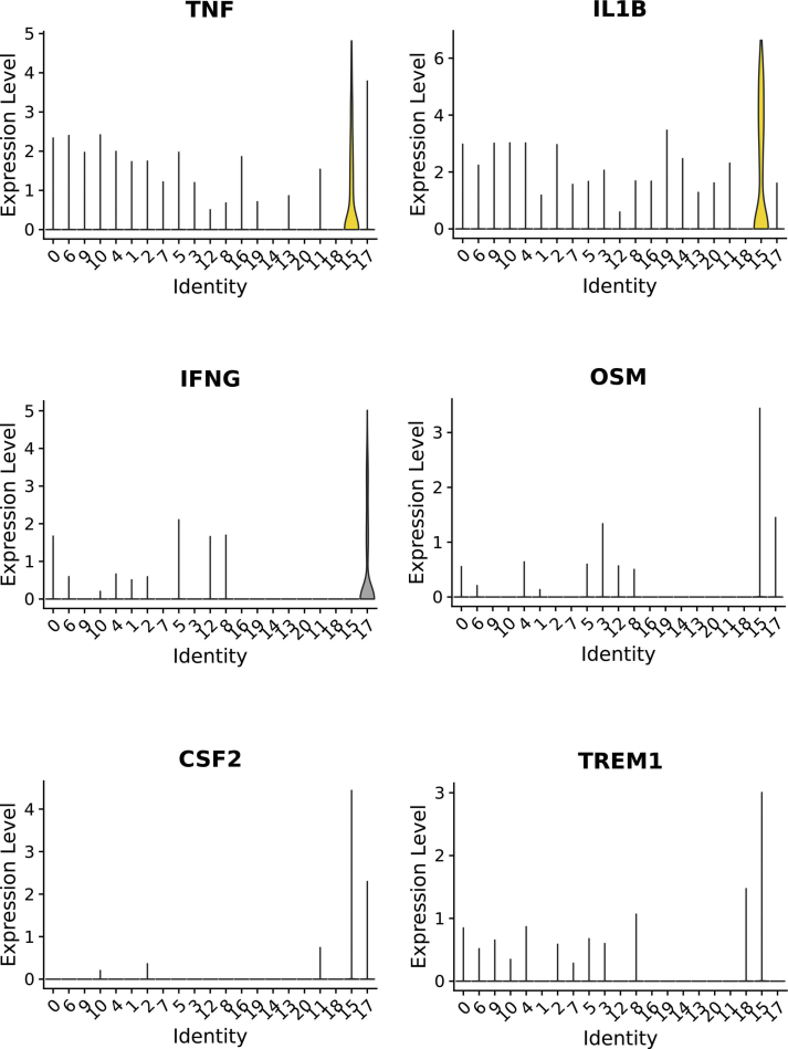
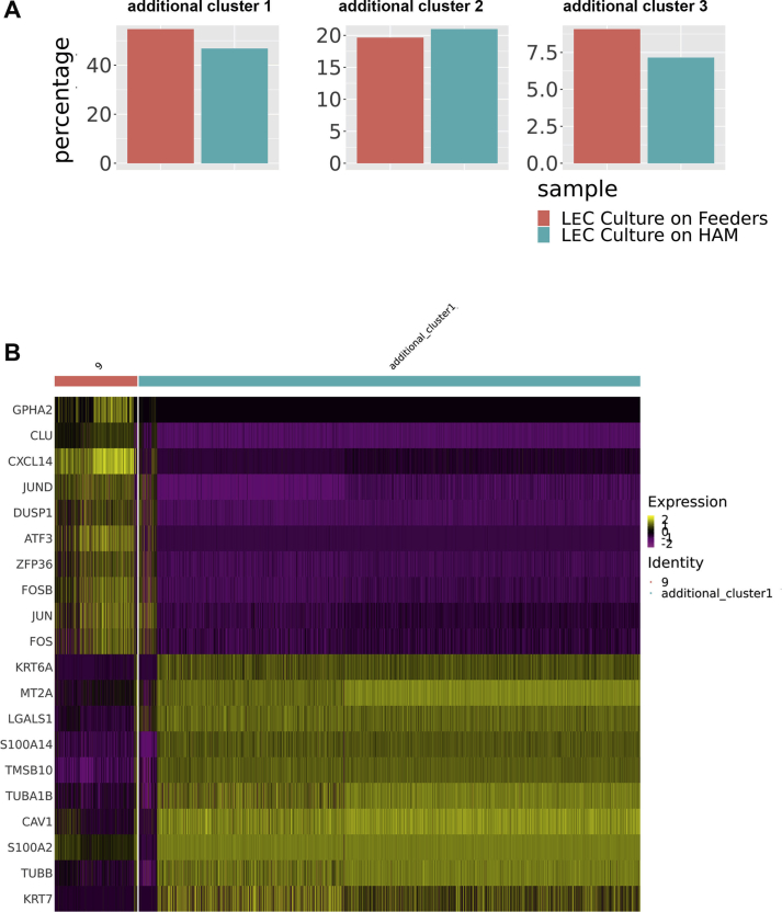
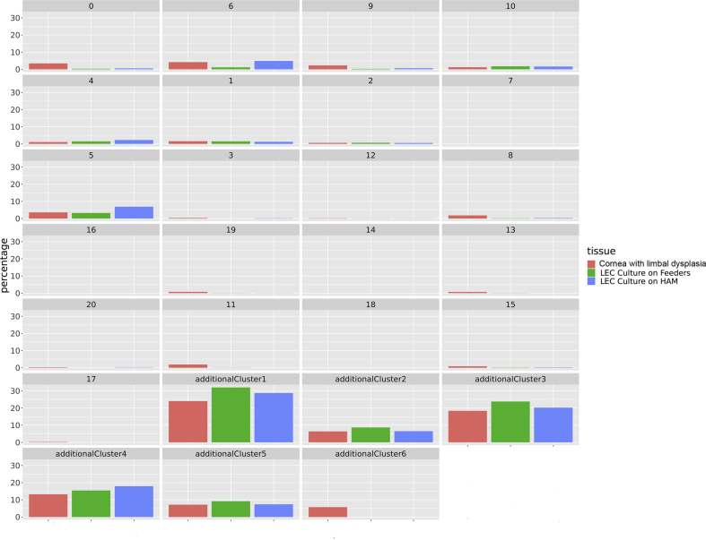
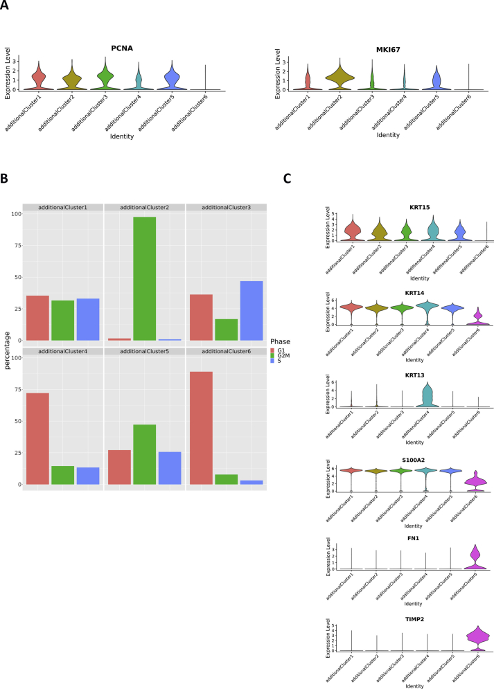
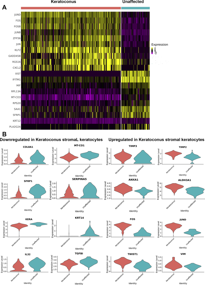
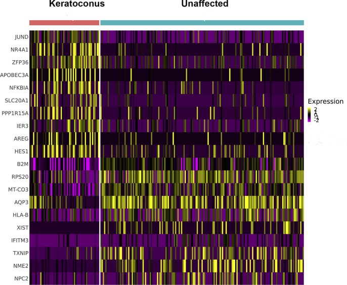
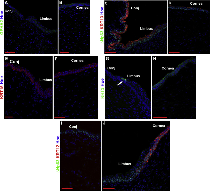
scRNA-Seq analysis of 21,343 cells from four adult human corneas and adjacent conjunctivas revealed the presence of 21 cell clusters, representing the progenitor and differentiated cells in all layers of cornea and conjunctiva as well as immune cells, melanocytes, fibroblasts, and blood/lymphatic vessels. A small cell cluster with high expression of limbal progenitor cell (LPC) markers was identified and shown via pseudotime analysis to give rise to five other cell types representing all the subtypes of differentiated limbal and corneal epithelial cells. A novel putative LPCs surface marker, GPHA2, expressed on the surface of 0.41% ± 0.21 of the cultured LECs, was identified, based on predominant expression in the limbal crypts of adult and developing cornea and RNAi validation in cultured LECs. Combining scRNA- and ATAC-Seq analyses, we identified multiple upstream regulators for LPCs and demonstrated a close interaction between the immune cells and limbal progenitor cells. RNA-Seq analysis indicated the loss of GPHA2 expression and acquisition of proliferative limbal basal epithelial cell markers during ex vivo LEC expansion, independently of the culture method used. Extending the single cell analyses to keratoconus, we were able to reveal activation of collagenase in the corneal stroma and a reduced pool of limbal suprabasal cells as two key changes underlying the disease phenotype. Single cell RNA-Seq of 89,897 cells obtained from embryonic and fetal cornea indicated that during development, the conjunctival epithelium is the first to be specified from the ocular surface epithelium, followed by the corneal epithelium and the establishment of LPCs, which predate the formation of limbal niche by a few weeks.
Our scRNA-and ATAC-Seq data of developing and adult cornea in steady state and disease conditions provide a unique resource for defining genes/pathways that can lead to improvement in ex vivo LPCs expansion, stem cell differentiation methods and better understanding and treatment of ocular surface disorders.
对关键的胚胎、胎儿和成人阶段的单细胞进行分析,以生成从发育到成年的所有角膜和邻近结膜细胞类型的综合单细胞图谱。
从 10 至 21 孕周的 4 个成人和 17 个胚胎和胎儿角膜中分离出单细胞,并使用 10x Genomics 平台进行单细胞 RNA 和/或 ATAC-Seq 分析。使用统一流形逼近和投影(UMAP)对这些细胞进行嵌入,并使用 Seurat 基于图的聚类进行聚类。基于标记基因表达、生物信息学数据挖掘和免疫荧光(IF)分析来确定簇的身份。对培养的角膜缘上皮细胞(LEC)进行 RNA 干扰、IF、集落形成效率和克隆分析。
对来自 4 个人类成人角膜和邻近结膜的 21343 个细胞的 scRNA-Seq 分析显示,存在 21 个细胞簇,代表角膜和结膜所有层的祖细胞和分化细胞以及免疫细胞、黑素细胞、成纤维细胞和血液/淋巴管。鉴定出一个具有高表达角膜缘祖细胞(LPC)标志物的小细胞簇,并通过伪时间分析显示其可分化为代表所有分化的角膜缘和角膜上皮细胞亚型的另外 5 种细胞类型。基于在成人和发育中的角膜缘隐窝中的主要表达和在培养的 LEC 中的 RNAi 验证,鉴定出一种新型的潜在 LPC 表面标志物 GPHA2,其在 0.41%±0.21%的培养 LEC 表面表达。通过 scRNA-和 ATAC-Seq 分析的结合,我们确定了 LPC 的多个上游调节剂,并证明了免疫细胞与角膜缘祖细胞之间的密切相互作用。RNA-Seq 分析表明,在体外 LEC 扩增过程中,GPHA2 表达丧失和获得增殖性角膜缘基底上皮细胞标志物,而与所使用的培养方法无关。将单细胞分析扩展到圆锥角膜,我们能够揭示角膜基质中胶原酶的激活和角膜缘上基细胞数量的减少,这是该疾病表型的两个关键变化。从胚胎和胎儿角膜获得的 89897 个细胞的单细胞 RNA-Seq 表明,在发育过程中,结膜上皮首先从眼表面上皮特化而来,随后是角膜上皮和 LPC 的建立,而 LPC 的建立比角膜缘龛的形成早几周。
我们对稳态和疾病条件下发育中和成人角膜的 scRNA-和 ATAC-Seq 数据提供了一个独特的资源,用于定义可导致体外 LPC 扩增、干细胞分化方法的改善以及更好地理解和治疗眼表疾病的基因/途径。