Guder Petra, Scheungrab Max, Kohnert Peter, Kolyfetis Georgios, Wanner Gerhard, Heß Martin
Staatliches Museum Für Naturkunde Karlsruhe, Erbprinzenstraße 13, Karlsruhe, 76133, Germany.
Biozentrum LMU München, Großhaderner Straße 2, Planegg-Martinsried, 82152, Germany.
BMC Biol. 2025 May 19;23(1):137. doi: 10.1186/s12915-025-02242-7.
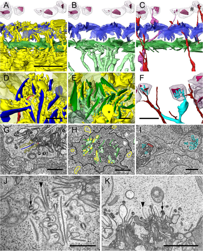
Block-face scanning electron microscopy has opened a new era of connectomics research, in which it is possible to make dense reconstructions of all cells in a clipping of a neuronal network, such as the retina, resolving synaptic contacts. Anchovies, exceptionally abundant marine teleosts, have retinae with regions for triple cone-based color vision and a region with specialized cone photoreceptors, so-called polycones, made of long and short cones with axially oriented outer segment lamellae for polarization contrast vision. This modality, discovered in the 1970s, is unique in vertebrates, but the neural wiring for contrast generation in deeper retinal layers is unknown so far.
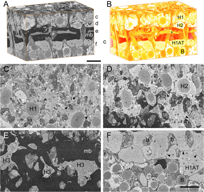
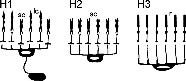
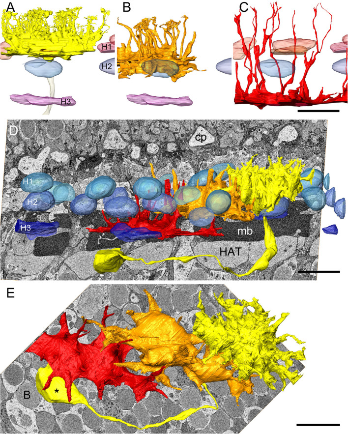
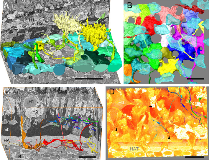
To elucidate the retinal connectomics of the European anchovy Engraulis encrasicolus (Linnaeus, 1758), in a first project, we investigated the shapes and cone-specific wiring rules of 3 horizontal cell types using volume electron microscopy and subsequent computer-aided reconstruction: H1 cells contact both cone types of the polycone, H2 cells contact only the short cones, and H3 cells are exclusively connected to rods. In addition, a distinctive double band of Müller fibers and a layer of H1 axon terminals were structurally clarified.
The findings suggest that (1) the monochromatic polarization contrast system based on fine structure specializations in the outer retina is connected to an inherited (bichromatic) color contrast mechanism in the inner retina, (2) the anchovy polycones arose from red (now long) and green (now short) cones, and (3) the blue single cones disappeared in the relevant retinal region.
块面扫描电子显微镜开启了连接组学研究的新时代,在这个时代,可以对神经网络切片(如视网膜)中的所有细胞进行密集重建,解析突触连接。凤尾鱼是数量异常丰富的海洋硬骨鱼,其视网膜具有基于三锥细胞的颜色视觉区域和一个具有特殊锥体细胞感受器的区域,即所谓的多锥体细胞,由长锥体细胞和短锥体细胞组成,其外段薄片呈轴向排列用于偏振对比视觉。这种模式在20世纪70年代被发现,在脊椎动物中是独一无二的,但迄今为止,视网膜深层用于产生对比的神经连接尚不清楚。
为了阐明欧洲凤尾鱼(Engraulis encrasicolus,林奈,1758)的视网膜连接组学,在第一个项目中,我们使用体积电子显微镜和随后的计算机辅助重建研究了3种水平细胞类型(H1细胞、H2细胞和H3细胞)的形状和特定于锥体细胞的连接规则:H1细胞与多锥体细胞的两种锥体细胞类型都有接触,H2细胞只与短锥体细胞接触,H3细胞仅与视杆细胞相连。此外,还在结构上明确了一条独特的米勒纤维双带和一层H1轴突终末。
研究结果表明:(1)基于视网膜外层精细结构特化的单色偏振对比系统与视网膜内层遗传的(双色)颜色对比机制相连;(2)凤尾鱼中的多锥体细胞由红色(现在的长锥体细胞)和绿色(现在的短锥体细胞)锥体细胞进化而来;(3)蓝色单锥体细胞在相关视网膜区域消失。