Ferraro Stefania, Demichelis Greta, Medina Carrion Jean Paul, Liu Dan, Becker Benjamin, Maes Michael, Fedeli Davide, Ciullo Giuseppe, Usai Susanna, Grisoli Marina, Chiapparini Luisa, Cecchini Proietti Alberto, Giani Luca, Nigri Anna, Leone Massimo
Sichuan Provincial Center for Mental Health, Sichuan Provincial People's Hospital, School of Medicine, University of Electronic Science and Technology of China, Chengdu, 610072, China.
School of Life Science and Technology, University of Electronic Science and Technology of China, Chengdu, China.
J Headache Pain. 2025 May 19;26(1):121. doi: 10.1186/s10194-025-02017-z.
This study aimed to identify mesocorticolimbic functional abnormalities in cluster headache (CH) patients, disentangling the roles of chronification and affective symptoms.
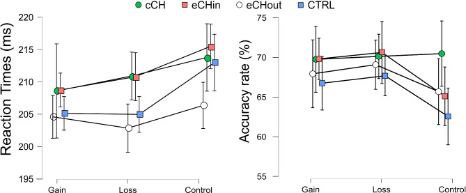
Using the monetary incentive delay fMRI task to directly engage these pathways, we investigated functional alterations in key regions of this network in chronic (n = 23) and episodic CH patients (n = 49) compared to a control group (n = 32). After processing the fMRI data, we extracted beta values from selected regions and for contrasts of interest and entered them into logistic regression models adjusted for potential confounders (such as depressive and anxiety symptoms and smoking habit) to test their association with the diagnoses (chronic CH and control subjects, episodic CH and control subjects).
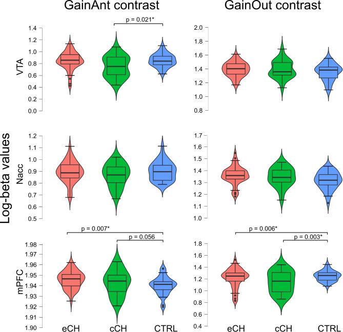
Results showed that chronic CH patients exhibited reduced ventral tegmental area (VTA) activity and a tendency towards significance (p = 0.056) for an increased medial prefrontal cortex (mPFC) responsiveness during reward anticipation, alongside a significant decrease in mPFC activity during reward outcomes. Episodic patients displayed abnormal mPFC activity across both reward phases, but coupled with intact VTA responses. Importantly, these functional abnormalities were not correlated to depressive and anxiety symptoms and smoking habits.
These findings suggest that chronic CH patients experience an imbalance in the VTA-mPFC pathway, while episodic patients may show early signs of this emerging dysfunction. Moreover, the observed reward processing alterations seem distinct from those associated with affective disorders, possibly highlighting unique mechanisms underlying the pathophysiology of CH.
本研究旨在确定丛集性头痛(CH)患者中脑皮质边缘系统的功能异常,厘清病程慢性化和情感症状的作用。
我们使用金钱激励延迟功能磁共振成像任务直接激活这些神经通路,调查了慢性丛集性头痛患者(n = 23)和发作性丛集性头痛患者(n = 49)与对照组(n = 32)相比,该神经网络关键区域的功能改变。在处理功能磁共振成像数据后,我们从选定区域提取β值并用于感兴趣的对比,然后将其输入经潜在混杂因素(如抑郁和焦虑症状以及吸烟习惯)校正的逻辑回归模型,以测试它们与诊断(慢性丛集性头痛患者与对照组、发作性丛集性头痛患者与对照组)的关联。
结果显示,慢性丛集性头痛患者在奖励预期期间腹侧被盖区(VTA)活动减少,内侧前额叶皮质(mPFC)反应性增加有显著趋势(p = 0.056),同时在奖励结果期间mPFC活动显著降低。发作性丛集性头痛患者在两个奖励阶段均表现出mPFC活动异常,但VTA反应正常。重要的是,这些功能异常与抑郁和焦虑症状以及吸烟习惯无关。
这些发现表明,慢性丛集性头痛患者在VTA - mPFC通路存在失衡,而发作性丛集性头痛患者可能显示出这种新出现功能障碍的早期迹象。此外,观察到的奖励处理改变似乎与情感障碍相关的改变不同,这可能突出了丛集性头痛病理生理学的独特机制。