• 文献检索
  • 文档翻译
  • 深度研究
  • 学术资讯
  • Suppr Zotero 插件Zotero 插件
  • 邀请有礼
  • 套餐&价格
  • 历史记录
应用&插件
Suppr Zotero 插件Zotero 插件浏览器插件Mac 客户端Windows 客户端微信小程序
定价
高级版会员购买积分包购买API积分包
服务
文献检索文档翻译深度研究API 文档MCP 服务
关于我们
关于 Suppr公司介绍联系我们用户协议隐私条款
关注我们

Suppr 超能文献

核心技术专利:CN118964589B侵权必究
粤ICP备2023148730 号-1Suppr @ 2026

文献检索

告别复杂PubMed语法,用中文像聊天一样搜索,搜遍4000万医学文献。AI智能推荐,让科研检索更轻松。

立即免费搜索

文件翻译

保留排版,准确专业,支持PDF/Word/PPT等文件格式,支持 12+语言互译。

免费翻译文档

深度研究

AI帮你快速写综述,25分钟生成高质量综述,智能提取关键信息,辅助科研写作。

立即免费体验

卵巢纤维瘤-卵泡膜细胞瘤组不同组织病理学亚型的传统超声和超声造影检查结果

Findings on conventional ultrasonography and contrast-enhanced ultrasonography in different histopathological subtypes of ovarian thecoma-fibroma group.

作者信息

Jiang Yuemingming, Ding Yanhua, Guo Minhua, Yu Yue, Duan Hongpeng, Zhang Shengmin

机构信息

Department of Ultrasonography, The First Affiliated Hospital of Ningbo University, 51#, Liuting Road, Ningbo, Zhejiang, 315010, China.

Department of Ultrasonography, Hangzhou Women's Hospital, Hangzhou, Zhejiang, China.

出版信息

BMC Med Imaging. 2025 May 20;25(1):175. doi: 10.1186/s12880-025-01693-2.

DOI:10.1186/s12880-025-01693-2
PMID:40394597
原文链接:https://pmc.ncbi.nlm.nih.gov/articles/PMC12090646/
Abstract

BACKGROUND

Ovarian thecoma-fibroma group (OTFG) is an unusual type of ovarian cancer with three histopathologic subtypes, but their features on ultrasonography are still poorly understood. This study evaluated the features of different histopathological subtypes of OTFG on conventional ultrasonography and contrast-enhanced ultrasonography (CEUS).

METHODS

This retrospective study enrolled sixty-nine women with pathologically confirmed OTFG who underwent preoperative CEUS. The characteristics of OTFG on conventional ultrasonography and CEUS, clinical manifestations, and laboratory findings were compared among subtypes.

RESULTS

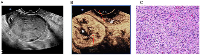
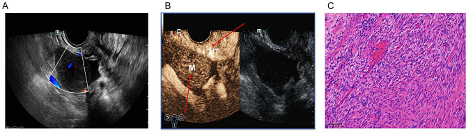
Fourteen patients were diagnosed with fibroma, fifty-one with thecofibroma, and four with thecoma. Although 69% of patients were post-menopausal, thecoma patients were significantly younger than those in other two groups. Laboratory examination revealed 21.7% (15/69) of patients had high carbohydrate antigen 125 (CA-125) level. On conventional ultrasonography, 72.5% (50/69) masses showed solid type, 24.6% (17/69) showed mixed cystic-solid type, and only 2.9% (2/69) showed cystic type. On CEUS, 50% (2/4) of thecoma lesions were rapid enhancement, 58.8% (30/51) of thecofibroma lesions and 78.6% (11/14) of fibroma lesions showed slow enhancement, 75% (3/4) of thecoma lesions showed isoenhancement during the descent process, and only 13.75% (7/51) of thecofibroma lesions and 7.1% (1/14) of fibroma lesions showed isoenhancement during the descent. they varied significantly among different histopathological subtypes.

CONCLUSIONS

The majority of OTFG is solid-like on conventional ultrasonography. Menopause is an important factor related to the subtype of OTFG. In postmenopausal patients with solid adnexal masses, slow hypoenhancement on CEUS is an important feature of fibroma. In premenopausal patients with solid or mixed cystic-solid adnexal masses, thecoma may be considered when rapid hyperenhancement, and isoenhancement or hypoenhancement during descent are observed on CEUS.

CLINICAL TRIAL NUMBER

Not applicable.

摘要

背景

卵巢纤维瘤-卵泡膜细胞瘤组(OTFG)是一种不常见的卵巢癌类型,有三种组织病理学亚型,但其超声特征仍了解不足。本研究评估了OTFG不同组织病理学亚型在传统超声和超声造影(CEUS)上的特征。

方法

本回顾性研究纳入了69例经病理证实为OTFG且术前行CEUS检查的女性患者。比较了各亚型OTFG在传统超声和CEUS上的特征、临床表现及实验室检查结果。

结果

14例患者诊断为纤维瘤,51例为纤维卵泡膜瘤,4例为卵泡膜瘤。尽管69%的患者已绝经,但卵泡膜瘤患者明显比其他两组患者年轻。实验室检查显示21.7%(15/69)的患者糖类抗原125(CA-125)水平升高。在传统超声上,72.5%(50/69)的肿块为实性,24.6%(17/69)为囊实性混合,仅2.9%(2/69)为囊性。在CEUS上,50%(2/4)的卵泡膜瘤病变呈快速增强,58.8%(30/51)的纤维卵泡膜瘤病变和78.6%(11/14)的纤维瘤病变呈缓慢增强,75%(3/4)的卵泡膜瘤病变在消退过程中呈等增强,而仅13.75%(7/51)的纤维卵泡膜瘤病变和7.1%(1/14)的纤维瘤病变在消退过程中呈等增强。不同组织病理学亚型之间差异显著。

结论

大多数OTFG在传统超声上呈类实性。绝经是与OTFG亚型相关的重要因素。在绝经后附件实性肿块患者中,CEUS上缓慢低增强是纤维瘤的重要特征。在绝经前附件实性或囊实性混合肿块患者中,当CEUS显示快速高增强、消退过程中等增强或低增强时,可能考虑卵泡膜瘤。

临床试验编号

不适用。

https://cdn.ncbi.nlm.nih.gov/pmc/blobs/6b6c/12090646/ed9d308a990a/12880_2025_1693_Fig3_HTML.jpg
https://cdn.ncbi.nlm.nih.gov/pmc/blobs/6b6c/12090646/25828f9b3f08/12880_2025_1693_Fig1_HTML.jpg
https://cdn.ncbi.nlm.nih.gov/pmc/blobs/6b6c/12090646/ecfcd99c20ee/12880_2025_1693_Fig2_HTML.jpg
https://cdn.ncbi.nlm.nih.gov/pmc/blobs/6b6c/12090646/ed9d308a990a/12880_2025_1693_Fig3_HTML.jpg
https://cdn.ncbi.nlm.nih.gov/pmc/blobs/6b6c/12090646/25828f9b3f08/12880_2025_1693_Fig1_HTML.jpg
https://cdn.ncbi.nlm.nih.gov/pmc/blobs/6b6c/12090646/ecfcd99c20ee/12880_2025_1693_Fig2_HTML.jpg
https://cdn.ncbi.nlm.nih.gov/pmc/blobs/6b6c/12090646/ed9d308a990a/12880_2025_1693_Fig3_HTML.jpg

相似文献

1
Findings on conventional ultrasonography and contrast-enhanced ultrasonography in different histopathological subtypes of ovarian thecoma-fibroma group.卵巢纤维瘤-卵泡膜细胞瘤组不同组织病理学亚型的传统超声和超声造影检查结果
BMC Med Imaging. 2025 May 20;25(1):175. doi: 10.1186/s12880-025-01693-2.
2
Ovarian thecoma-fibroma groups: clinical and sonographic features with pathological comparison.卵巢纤维瘤-卵泡膜细胞瘤组:临床及超声特征与病理对照
J Ovarian Res. 2016 Nov 22;9(1):81. doi: 10.1186/s13048-016-0291-2.
3
CT diagnosis in the thecoma-fibroma group of the ovarian stromal tumors.卵巢间质肿瘤中卵泡膜细胞瘤-纤维瘤组的CT诊断
Cell Biochem Biophys. 2015 Mar;71(2):937-43. doi: 10.1007/s12013-014-0288-7.
4
Conventional ultrasound and high-frame-rate contrast-enhanced ultrasound characteristics of ovarian thecoma-fibroma groups.卵巢纤维瘤-卵泡膜细胞瘤组的传统超声及高帧率超声造影特征
Quant Imaging Med Surg. 2025 May 1;15(5):3875-3890. doi: 10.21037/qims-24-2200. Epub 2025 Apr 17.
5
Prediction of benignity of solid adnexal masses.附件实性肿块良恶性的预测。
Arch Gynecol Obstet. 2012 Mar;285(3):721-6. doi: 10.1007/s00404-011-2062-z. Epub 2011 Aug 20.
6
Pelvic effusion in patients with ovarian thecoma-fibroma associated with the tumor size and plaste CA-125 level: A retrospective magnetic resonance imaging study.卵巢卵泡膜细胞瘤-纤维瘤患者的盆腔积液与肿瘤大小和 plaste CA-125 水平的关系:一项回顾性磁共振成像研究。
Clin Imaging. 2022 Jan;81:62-66. doi: 10.1016/j.clinimag.2021.06.007. Epub 2021 Aug 3.
7
Ovarian fibroma/fibrothecoma: retrospective cohort study shows limited value of risk of malignancy index score.卵巢纤维瘤/纤维卵泡膜瘤:回顾性队列研究显示恶性风险指数评分价值有限。
Aust N Z J Obstet Gynaecol. 2013 Jun;53(3):287-92. doi: 10.1111/ajo.12090. Epub 2013 Apr 23.
8
[Comparision of sonographic findings and pathological features in ovarian thecoma].卵巢卵泡膜细胞瘤超声表现与病理特征的比较
Zhonghua Zhong Liu Za Zhi. 2008 Jun;30(6):473-5.
9
Imaging in gynecological disease (5): clinical and ultrasound characteristics in fibroma and fibrothecoma of the ovary.妇科疾病影像学(五):卵巢纤维瘤和纤维卵泡膜细胞瘤的临床及超声特征。
Ultrasound Obstet Gynecol. 2009 Aug;34(2):188-95. doi: 10.1002/uog.6394.
10
Magnetic resonance imaging features of ovarian fibroma, fibrothecoma, and thecoma.卵巢纤维瘤、纤维卵泡膜瘤和卵泡膜瘤的磁共振成像特征。
Abdom Imaging. 2015 Jun;40(5):1263-72. doi: 10.1007/s00261-014-0257-z.

本文引用的文献

1
High altitudes, deeper insights: multicenter cardiovascular magnetic resonance study on hypertrophic cardiomyopathy.高海拔地区,深入洞察:肥厚型心肌病的多中心心血管磁共振研究
Eur Radiol. 2024 Dec 31. doi: 10.1007/s00330-024-11305-2.
2
Deep learning model based on contrast-enhanced ultrasound for predicting vessels encapsulating tumor clusters in hepatocellular carcinoma.基于超声造影的深度学习模型用于预测肝细胞癌中包裹肿瘤结节的血管情况
Eur Radiol. 2025 Feb;35(2):989-1000. doi: 10.1007/s00330-024-10985-0. Epub 2024 Jul 27.
3
The Current Role of Contrast-Enhanced Ultrasound (CEUS) in the Diagnosis and Staging of Bladder Cancer: A Review of the Available Literature.
超声造影(CEUS)在膀胱癌诊断和分期中的当前作用:现有文献综述
Life (Basel). 2024 Jul 9;14(7):857. doi: 10.3390/life14070857.
4
Efficacy of color Doppler ultrasound and contrast-enhanced ultrasound in identifying vascular invasion in pancreatic ductal adenocarcinoma.彩色多普勒超声与超声造影在鉴别胰腺导管腺癌血管侵犯中的效能
Insights Imaging. 2024 Jul 25;15(1):181. doi: 10.1186/s13244-024-01779-5.
5
Contrast-Enhanced Ultrasound in the Diagnosis of Solid Renal Lesions.超声造影在肾实性病变诊断中的应用
J Clin Med. 2024 Jun 29;13(13):3821. doi: 10.3390/jcm13133821.
6
Intraoperative frozen section evaluation of ovarian sex cord-stromal tumours and their mimics: a study of 121 cases with emphasis on potential diagnostic pitfalls.卵巢性索-间质肿瘤及其类似物的术中冰冻切片评估:121 例病例研究,重点关注潜在的诊断陷阱。
Pathology. 2024 Oct;56(6):842-853. doi: 10.1016/j.pathol.2024.04.007. Epub 2024 Jun 20.
7
The value of contrast-enhanced ultrasound in differentiating benign from malignant retroperitoneal masses.增强超声在鉴别腹膜后良恶性肿块中的价值。
Eur J Radiol. 2024 Sep;178:111596. doi: 10.1016/j.ejrad.2024.111596. Epub 2024 Jun 29.
8
Diagnostic performance of contrast-enhanced ultrasound (CEUS) combined with Ovarian-Adnexal Reporting and Data System (O-RADS) ultrasound risk stratification for adnexal masses: a systematic review and meta-analysis.超声造影(CEUS)联合卵巢附件报告和数据系统(O-RADS)超声风险分层对附件包块的诊断性能:一项系统评价和荟萃分析
Clin Radiol. 2024 Sep;79(9):e1167-e1175. doi: 10.1016/j.crad.2024.05.021. Epub 2024 Jun 7.
9
A modified CEUS risk stratification model for adnexal masses with solid components: prospective multicenter study and risk adjustment.改良的附件区实性肿块超声造影风险分层模型:前瞻性多中心研究和风险调整。
Eur Radiol. 2024 Sep;34(9):5978-5988. doi: 10.1007/s00330-024-10639-1. Epub 2024 Feb 19.
10
Ultrasonographic manifestations and misdiagnosis analysis of ovarian leiomyoma.卵巢平滑肌瘤的超声表现及误诊分析
Front Med (Lausanne). 2023 Dec 27;10:1289522. doi: 10.3389/fmed.2023.1289522. eCollection 2023.