Santarelli Valerio, Rosati Davide, Canale Vittorio, Salciccia Stefano, Di Lascio Giovanni, Bevilacqua Giulio, Tufano Antonio, Sciarra Alessandro, Cantisani Vito, Franco Giorgio, Moriconi Martina, Di Pierro Giovanni Battista
Department of Maternal-Infant and Urological Sciences, "Sapienza" Rome University, Policlinico Umberto I Hospital, 00185 Rome, Italy.
Department of Radiology, Oncology and Pathology, University La Sapienza of Rome, 00185 Roma, Italy.
Life (Basel). 2024 Jul 9;14(7):857. doi: 10.3390/life14070857.
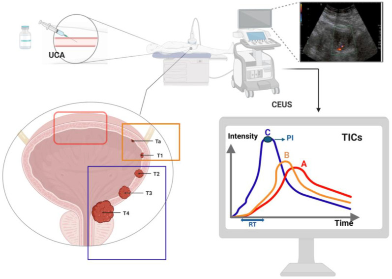
Contrast-enhanced ultrasound (CEUS) is an advanced imaging technique that integrates conventional US with the intravenous injection of specific US contrast agents (UCAs), combining the non-invasiveness of US with the higher accuracy of contrast-enhanced imaging. In contrast with magnetic resonance imaging (MRI), computed tomography (CT) and cystoscopy, CEUS has few contraindications, and UCAs are non-nephrotoxic agents that can be safely used in patients with kidney failure. CEUS is a well-established method for the detection of liver lesions and for echocardiography, and its indications are expanding. The updated 2018 WFUMB-EFSUMB guidelines have added the urinary bladder under non-hepatic applications of CEUS. The technique is able to distinguish between benign tissue, such as clots or hematoma, and malignant lesions by perfusing the mass with contrast agent. Thanks to the different perfusion rates of the various layers of the bladder wall, CEUS is also able to predict tumor invasion depth and stage. Despite that, current urological guidelines do not include CEUS as a plausible imaging technique for bladder urothelial carcinoma. The main reason for this omission might be the presence of scarce randomized evidence and the absence of large validated series. In this review, we describe the rationale behind the use of CEUS in bladder cancer and the added value of this imaging technique in the detection and staging of bladder lesions. In addition, we researched the available literature on the topic and then described the results of randomized clinical trials and a meta-analysis investigating the accuracy of CEUS in bladder cancer diagnosis and staging. The reported studies show that CEUS is a highly accurate diagnostic and staging tool for BC, reaching levels of specificity and sensitivity in differentiating between Ta-T1, or low-grade BC, and T2, or high-grade BC, that are comparable to those shown by the reference standard methods. Nonetheless, several limitations were found and are highlighted in this review. The aim of this study is to further validate and promote the use of CEUS as a quick, economic and effective diagnostic tool for this high-impact disease.
超声造影(CEUS)是一种先进的成像技术,它将传统超声与静脉注射特定的超声造影剂(UCA)相结合,将超声的非侵入性与造影增强成像的更高准确性结合起来。与磁共振成像(MRI)、计算机断层扫描(CT)和膀胱镜检查相比,CEUS的禁忌症很少,并且UCA是非肾毒性药物,可安全用于肾衰竭患者。CEUS是一种成熟的检测肝脏病变和进行超声心动图检查的方法,其应用范围正在不断扩大。2018年更新的WFUMB-EFSUMB指南在CEUS的非肝脏应用中增加了膀胱。该技术能够通过向肿块灌注造影剂来区分良性组织,如血凝块或血肿,以及恶性病变。由于膀胱壁各层的灌注速率不同,CEUS还能够预测肿瘤的浸润深度和分期。尽管如此,目前的泌尿外科指南并未将CEUS列为膀胱尿路上皮癌的一种可行成像技术。这一遗漏的主要原因可能是缺乏随机证据以及没有大量经过验证的系列研究。在本综述中,我们描述了在膀胱癌中使用CEUS的基本原理以及这种成像技术在膀胱病变检测和分期中的附加价值。此外,我们研究了关于该主题的现有文献,然后描述了随机临床试验和一项荟萃分析的结果,该荟萃分析调查了CEUS在膀胱癌诊断和分期中的准确性。报道的研究表明,CEUS是一种用于膀胱癌的高度准确的诊断和分期工具,在区分Ta-T1期或低级别膀胱癌与T2期或高级别膀胱癌方面达到的特异性和敏感性水平与参考标准方法相当。尽管如此,本综述中发现并强调了几个局限性。本研究的目的是进一步验证并推广将CEUS作为这种高影响疾病的快速、经济且有效的诊断工具。