• 文献检索
  • 文档翻译
  • 深度研究
  • 学术资讯
  • Suppr Zotero 插件Zotero 插件
  • 邀请有礼
  • 套餐&价格
  • 历史记录
应用&插件
Suppr Zotero 插件Zotero 插件浏览器插件Mac 客户端Windows 客户端微信小程序
定价
高级版会员购买积分包购买API积分包
服务
文献检索文档翻译深度研究API 文档MCP 服务
关于我们
关于 Suppr公司介绍联系我们用户协议隐私条款
关注我们

Suppr 超能文献

核心技术专利:CN118964589B侵权必究
粤ICP备2023148730 号-1Suppr @ 2026

文献检索

告别复杂PubMed语法,用中文像聊天一样搜索,搜遍4000万医学文献。AI智能推荐,让科研检索更轻松。

立即免费搜索

文件翻译

保留排版,准确专业,支持PDF/Word/PPT等文件格式,支持 12+语言互译。

免费翻译文档

深度研究

AI帮你快速写综述,25分钟生成高质量综述,智能提取关键信息,辅助科研写作。

立即免费体验

一例伴有内镜形态学改变的胃非典型脂肪瘤样肿瘤/高分化脂肪肉瘤

A Case of Gastric Atypical Lipomatous Tumor/Well-Differentiated Liposarcoma With Endoscopic Morphological Changes.

作者信息

Omote Rika, Omote Shizuma, Sonobe Hiroshi, Hamano Ryosuke, Toyokawa Tatsuya, Otsuka Shinya, Tanaka Takehiro, Yanai Hiroyuki, Inagaki Masaru, Yamamoto Hidetaka

机构信息

Department of Diagnostic Pathology NHO Fukuyama Medical Center Hiroshima Japan.

Department of Internal Medicine Fukuyama Minami Hospital Hiroshima Japan.

出版信息

DEN Open. 2025 May 22;6(1):e70146. doi: 10.1002/deo2.70146. eCollection 2026 Apr.

DOI:10.1002/deo2.70146
PMID:40416586
原文链接:https://pmc.ncbi.nlm.nih.gov/articles/PMC12098964/
Abstract

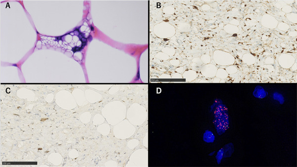
Atypical lipomatous tumor/well-differentiated liposarcoma is a locally aggressive mesenchymal neoplasm composed of adipocytes and stromal cells. Gastric cases are exceedingly rare, and their malignant potential remains unclear. We report a case of a woman in her 60s who was found to have multiple submucosal tumor-like lesions of the stomach. Over time, the tumors increased in size, requiring a laparoscopic partial gastrectomy. Histological examination revealed a tumor composed of both fatty tissue and fibrous stroma with nuclear atypia. Immunohistochemistry showed positivity for CDK4 and MDM2, and fluorescence in situ hybridization confirmed amplification, leading to a diagnosis of atypical lipomatous tumor/well-differentiated liposarcoma. This case presented an unusual gastric manifestation, with multiple submucosal tumor-like lesions on endoscopy and exhibiting progressive morphological changes over several years.

摘要

非典型脂肪瘤性肿瘤/高分化脂肪肉瘤是一种由脂肪细胞和基质细胞组成的具有局部侵袭性的间叶性肿瘤。胃的病例极为罕见,其恶性潜能仍不明确。我们报告一例60多岁女性,发现其胃有多个黏膜下肿瘤样病变。随着时间推移,肿瘤体积增大,需行腹腔镜部分胃切除术。组织学检查显示肿瘤由脂肪组织和纤维性基质组成,伴有核异型性。免疫组化显示CDK4和MDM2呈阳性,荧光原位杂交证实有扩增,从而诊断为非典型脂肪瘤性肿瘤/高分化脂肪肉瘤。该病例呈现出不寻常的胃部表现,内镜检查可见多个黏膜下肿瘤样病变,并在数年内呈现出渐进性形态变化。

https://cdn.ncbi.nlm.nih.gov/pmc/blobs/2436/12098964/3194cb1e925e/DEO2-6-e70146-g002.jpg
https://cdn.ncbi.nlm.nih.gov/pmc/blobs/2436/12098964/b9cc64ed0f01/DEO2-6-e70146-g003.jpg
https://cdn.ncbi.nlm.nih.gov/pmc/blobs/2436/12098964/a5e37dab1ea2/DEO2-6-e70146-g004.jpg
https://cdn.ncbi.nlm.nih.gov/pmc/blobs/2436/12098964/b1e8f43148ac/DEO2-6-e70146-g001.jpg
https://cdn.ncbi.nlm.nih.gov/pmc/blobs/2436/12098964/3194cb1e925e/DEO2-6-e70146-g002.jpg

相似文献

1
A Case of Gastric Atypical Lipomatous Tumor/Well-Differentiated Liposarcoma With Endoscopic Morphological Changes.一例伴有内镜形态学改变的胃非典型脂肪瘤样肿瘤/高分化脂肪肉瘤
DEN Open. 2025 May 22;6(1):e70146. doi: 10.1002/deo2.70146. eCollection 2026 Apr.
2
Dysplastic Lipoma: A Distinctive Atypical Lipomatous Neoplasm With Anisocytosis, Focal Nuclear Atypia, p53 Overexpression, and a Lack of MDM2 Gene Amplification by FISH; A Report of 66 Cases Demonstrating Occasional Multifocality and a Rare Association With Retinoblastoma.发育不良性脂肪瘤:一种具有异形性、局灶性核异型、p53 过表达且 FISH 检测无 MDM2 基因扩增的独特非典型脂肪性肿瘤;66 例报告显示其偶有多发和罕见与视网膜母细胞瘤相关。
Am J Surg Pathol. 2018 Nov;42(11):1530-1540. doi: 10.1097/PAS.0000000000001129.
3
Well-differentiated spindle cell liposarcoma ('atypical spindle cell lipomatous tumor') does not belong to the spectrum of atypical lipomatous tumor but has a close relationship to spindle cell lipoma: clinicopathologic, immunohistochemical, and molecular analysis of six cases.分化良好的梭形细胞脂肪肉瘤(“非典型梭形细胞脂肪瘤性肿瘤”)不属于非典型脂肪瘤性肿瘤范畴,而与梭形细胞脂肪瘤密切相关:六例病例的临床病理、免疫组织化学和分子分析。
Mod Pathol. 2010 May;23(5):729-36. doi: 10.1038/modpathol.2010.66. Epub 2010 Mar 12.
4
Overdiagnosis of atypical lipomatous tumors/well-differentiated liposarcomas by morphological diagnosis using only HE stained specimens: a case-control study with MDM2/CDK4 immunostaining and MDM2/CDK4 fluorescence in situ hybridization.仅通过 HE 染色标本的形态学诊断对非典型性脂肪肉瘤/高分化脂肪肉瘤的过度诊断:一项使用 MDM2/CDK4 免疫组化和 MDM2/CDK4 荧光原位杂交的病例对照研究。
BMC Cancer. 2024 Nov 21;24(1):1437. doi: 10.1186/s12885-024-13215-4.
5
Carboxypeptidase M: a biomarker for the discrimination of well-differentiated liposarcoma from lipoma.羧肽酶 M:一种用于鉴别高分化脂肪肉瘤与脂肪瘤的生物标志物。
Mod Pathol. 2009 Dec;22(12):1541-7. doi: 10.1038/modpathol.2009.149. Epub 2009 Oct 9.
6
Lipomas of the Oral Cavity: Utility of MDM2 and CDK4 in Avoiding Overdiagnosis as Atypical Lipomatous Tumor.口腔脂肪瘤:MDM2和CDK4在避免将其误诊为非典型脂肪瘤性肿瘤中的作用
Head Neck Pathol. 2019 Jun;13(2):169-176. doi: 10.1007/s12105-018-0928-0. Epub 2018 May 10.
7
Detection of MDM2/CDK4 amplification in lipomatous soft tissue tumors from formalin-fixed, paraffin-embedded tissue: comparison of multiplex ligation-dependent probe amplification (MLPA) and fluorescence in situ hybridization (FISH).福尔马林固定、石蜡包埋组织中脂肪性软组织肿瘤MDM2/CDK4扩增的检测:多重连接依赖探针扩增(MLPA)与荧光原位杂交(FISH)的比较
Appl Immunohistochem Mol Morphol. 2015 Feb;23(2):126-33. doi: 10.1097/PDM.0000000000000041.
8
Atypical lipomatous tumor/well differentiated liposarcoma and related mimics with updates. When is molecular testing most cost-effective, necessary, and indicated?非典型性脂肪肿瘤/高分化脂肪肉瘤及相关模拟肿瘤的最新研究进展。何时进行分子检测最具成本效益、最必要和最有指示性?
Hum Pathol. 2024 May;147:82-91. doi: 10.1016/j.humpath.2023.12.005. Epub 2023 Dec 21.
9
Lessons learnt from MDM2 fluorescence in-situ hybridisation analysis of 439 mature lipomatous lesions with an emphasis on atypical lipomatous tumour/well-differentiated liposarcoma lacking cytological atypia.从439例成熟脂肪性病变的MDM2荧光原位杂交分析中获得的经验教训,重点关注缺乏细胞学异型性的非典型脂肪瘤性肿瘤/高分化脂肪肉瘤。
Histopathology. 2022 Jan;80(2):369-380. doi: 10.1111/his.14558. Epub 2021 Nov 11.
10
Sensitivity of MDM2 amplification and unexpected multiple faint alphoid 12 (alpha 12 satellite sequences) signals in atypical lipomatous tumor.在非典型性脂肪瘤样肿瘤中 MDM2 扩增的敏感性和意想不到的多个微弱的α12(α12 卫星序列)信号。
Mod Pathol. 2012 Oct;25(10):1384-96. doi: 10.1038/modpathol.2012.90. Epub 2012 Jun 15.

本文引用的文献

1
Cellular origin and clonal evolution of human dedifferentiated liposarcoma.人类去分化脂肪肉瘤的细胞起源和克隆进化。
Nat Commun. 2024 Sep 12;15(1):7941. doi: 10.1038/s41467-024-52067-1.
2
Liposarcoma of the stomach: Report of two cases and review of the literature.胃脂肪肉瘤:两例报告并文献复习。
World J Gastroenterol. 2018 Jul 7;24(25):2776-2784. doi: 10.3748/wjg.v24.i25.2776.
3
MDM2 and CDK4 Immunohistochemistry: Should It Be Used in Problematic Differentiated Lipomatous Tumors?: A New Perspective.MDM2和CDK4免疫组化:是否应用于疑难性分化型脂肪肿瘤?一种新观点。
Am J Surg Pathol. 2016 Dec;40(12):1647-1652. doi: 10.1097/PAS.0000000000000713.
4
Can MDM2 analytical tests performed on core needle biopsy be relied upon to diagnose well-differentiated liposarcoma?核心针活检上进行的 MDM2 分析检测能否可靠诊断为高分化脂肪肉瘤?
Mod Pathol. 2010 Oct;23(10):1301-6. doi: 10.1038/modpathol.2010.106. Epub 2010 May 21.
5
Detection of MDM2-CDK4 amplification by fluorescence in situ hybridization in 200 paraffin-embedded tumor samples: utility in diagnosing adipocytic lesions and comparison with immunohistochemistry and real-time PCR.通过荧光原位杂交技术检测200份石蜡包埋肿瘤样本中的MDM2-CDK4扩增:在诊断脂肪细胞性病变中的应用及与免疫组织化学和实时PCR的比较
Am J Surg Pathol. 2007 Oct;31(10):1476-89. doi: 10.1097/PAS.0b013e3180581fff.
6
Atypical lipomatous tumor, its variants, and its combined forms: a study of 61 cases, with a minimum follow-up of 10 years.非典型脂肪瘤性肿瘤及其变异型和联合形式:61例研究,最短随访10年。
Am J Surg Pathol. 2007 Jan;31(1):1-14. doi: 10.1097/01.pas.0000213406.95440.7a.
7
Dedifferentiated liposarcoma: a clinicopathological analysis of 155 cases with a proposal for an expanded definition of dedifferentiation.去分化脂肪肉瘤:155例临床病理分析及去分化扩展定义的提议
Am J Surg Pathol. 1997 Mar;21(3):271-81. doi: 10.1097/00000478-199703000-00002.