Fadda Gian Luca, Saibene Alberto Maria, Rustichelli Chiara, Nitro Letizia, Lentini Mario, Parisi Federica Maria, Cocuzza Salvatore, Cavallo Giovanni, De Corso Eugenio, Maniaci Antonino
Department of Otolaryngology, University of Turin, "San Luigi Gonzaga" Hospital, Regione Gonzole 10, Orbassano, 10043 Turin, Italy.
Otolaryngology Unit, Santi Paolo e Carlo Hospital, Department of Health Sciences, Università degli Studi di Milano, 20142 Milan, Italy.
J Clin Med. 2025 May 16;14(10):3508. doi: 10.3390/jcm14103508.
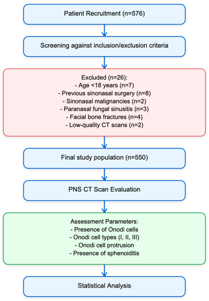
Sphenoiditis poses diagnostic and treatment challenges due to its complex anatomy and potential for serious complications. Anatomic variations, such as Onodi cells, could play a role in the onset and spreading of inflammation. The diagnosis and treatment of sphenoiditis can be more difficult if Onodi cells are present, especially due to their proximity to delicate vital tissues. : The purpose of this study was to look at the frequency, features, and relationship between Onodi cells and sphenoiditis. : This multicentric study comprised 550 people who received sinonasal CT imaging. The Thimmaiah classification was used to assess the presence and features of Onodi cells, and radiographic results were used to diagnose sphenoiditis. We conducted univariate and multivariate logistic regression to evaluate the relationships between sphenoiditis and Onodi cells. : The prevalence of Onodi cells was 32.40%, with a higher prevalence on the right side (18.40%) compared to the left side (8.40%). The multivariable analysis revealed a significant correlation between right-side Type II Onodi cells and a higher incidence of sphenoiditis (OR = 6.81, 95% CI: 1.14-38.97, = 0.029). In the univariable analysis (OR = 3.00, 95% CI: 1.15-6.96, = 0.015), but not in the multivariable analysis, the presence of Type I Onodi cells on the left side was significantly associated with sphenoiditis. : There may be a link between a higher incidence of sphenoiditis and the presence of Type II Onodi cells on the right side. In order to validate these findings and clarify the underlying processes of this connection, more prospective research is required.
由于蝶窦炎解剖结构复杂且有引发严重并发症的可能,因此其诊断和治疗颇具挑战。解剖变异,如Onodi气房,可能在炎症的发生和扩散中起作用。如果存在Onodi气房,蝶窦炎的诊断和治疗会更加困难,尤其是因为它们靠近脆弱的重要组织。本研究的目的是观察Onodi气房的发生率、特征以及与蝶窦炎的关系。这项多中心研究纳入了550例接受鼻窦CT成像的患者。采用Thimmaiah分类法评估Onodi气房的存在及特征,并利用影像学结果诊断蝶窦炎。我们进行了单变量和多变量逻辑回归分析,以评估蝶窦炎与Onodi气房之间的关系。Onodi气房的发生率为32.40%,右侧(18.40%)的发生率高于左侧(8.40%)。多变量分析显示,右侧II型Onodi气房与蝶窦炎较高的发病率之间存在显著相关性(OR = 6.81,95%CI:1.14 - 38.97,P = 0.029)。在单变量分析中(OR = 3.00,95%CI:1.15 - 6.96,P = 0.015),左侧I型Onodi气房的存在与蝶窦炎显著相关,但在多变量分析中并非如此。右侧II型Onodi气房的存在可能与蝶窦炎较高的发病率之间存在关联。为了验证这些发现并阐明这种关联的潜在机制,需要更多的前瞻性研究。