Helmi Zeena, Nori Wassan
Department of Obstetrics and Gynecology, Mustansiriyah University, Baghdad, Iraq*Correspondence: Wassan Nori. Email:
Qatar Med J. 2025 Jan 21;2025(1):24. doi: 10.5339/qmj.2025.24. eCollection 2025.
Endometrial osseous metaplasia (OM) is a rare condition characterized by the transformation of endometrial tissue into bone cells. Despite its rarity, OM remains a significant contributor to infertility. Although the underlying mechanism remains debatable, an association with previous abortions and curettage has been suggested.
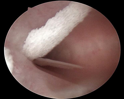
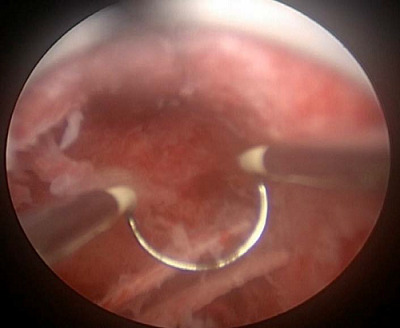
We present two cases of OM presented to the infertility clinic and discuss their similarities and discrepancies in presentation and risk factors. A transvaginal ultrasound raises suspicion about the diagnosis of OM with a hyperechoic mass and post-acoustic shadowing. An office hysteroscopy showed white, mesh-like bony sheets. Both cases underwent operative hysteroscopy to address surgical challenges, and the two cases were followed postoperatively for one year.
A comprehensive literature review examined various aspects of OM, including diagnosis, therapeutic options, outcomes, prognosis, and follow-up. Our aim was to raise awareness of this intriguing condition by providing up-to-date knowledge and emphasizing the central role of hysteroscopy in diagnosis and treatment. Here, we present two cases with the same complaint, infertility. Moreover, although the same treatment method was used in both cases, only one achieved pregnancy. This highlights that OM is a possible underlying cause of infertility, in addition to considering other factors that contribute to the overall clinical picture.
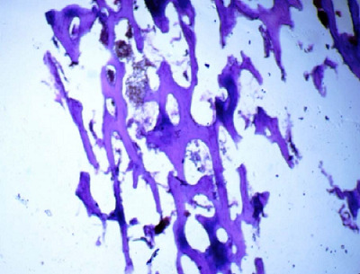
OM should be considered in the evaluation of infertility despite its rarity, especially with hyperechoic lesions and acoustic shadowing on ultrasound examination. Hysteroscopy is the gold standard for diagnosis and therapeutic approaches. A complete understanding of the reasons that trigger its growth is crucial. To rule out other differential diagnosis, a holistic evaluation of the patient's history, imaging, and histopathological examination is needed.
子宫内膜骨化生(OM)是一种罕见病症,其特征为子宫内膜组织转化为骨细胞。尽管罕见,但OM仍是导致不孕的一个重要因素。虽然其潜在机制仍存在争议,但已有研究表明它与既往流产和刮宫有关。
我们报告了两例在不孕不育诊所就诊的OM病例,并讨论了它们在临床表现和危险因素方面的异同。经阴道超声检查发现高回声团块及后方声影,提示可能为OM。门诊宫腔镜检查显示白色、网状的骨片。两例均接受了宫腔镜手术以应对手术挑战,并在术后随访一年。
通过全面的文献综述,研究了OM的各个方面,包括诊断、治疗选择、结局、预后及随访。我们的目的是通过提供最新知识并强调宫腔镜在诊断和治疗中的核心作用,提高对这种有趣病症的认识。在此,我们展示了两例主诉相同(不孕)的病例。此外,尽管两例采用了相同的治疗方法,但只有一例成功受孕。这突出表明,除了考虑导致整体临床情况的其他因素外,OM可能是不孕的潜在原因。
尽管OM罕见,但在评估不孕时应予以考虑,尤其是超声检查发现高回声病变及声影时。宫腔镜是诊断和治疗方法的金标准。全面了解触发其生长的原因至关重要。为排除其他鉴别诊断,需要对患者的病史、影像学和组织病理学检查进行全面评估。