Sankar Wudbhav N, Lee Julianna, Chong David, Hailer Yasmin D, Agrizzi de Angeli Luiz R, Yang Scott, Laine Jennifer, Kim Harry K W
Division of Orthopaedics, The Children's Hospital of Philadelphia, Philadelphia, PA, USA.
Department of Orthopaedic Surgery, University of Oklahoma, Oklahoma City, OK, USA.
J Pediatr Soc North Am. 2024 Feb 28;6:100019. doi: 10.1016/j.jposna.2024.100019. eCollection 2024 Feb.
Legg-Calvé-Perthes disease (LCPD) progresses through 4 stages characterized by unique radiographic features, and stage duration is recognized as an important prognostic factor. Newer perfusion magnetic resonance imaging (pMRI) allows for the evaluation of vascularity early in the disease process. This study aims to describe the relationship between global and regional perfusion patterns on early pMRI and the duration of Waldenström stages. A secondary aim was to verify the relationship between hypoperfusion and subsequent lateral pillar class.
Through a prospectively collected multicenter international cohort, patients with early LCPD (Waldenström Stage I) and pMRI were followed with serial radiographs at 3-month intervals for a minimum of 2 years. Epiphyseal hypoperfusion was quantified by HipVasc Software for the entire epiphysis and regional thirds of the femoral head. Waldenström stages and lateral pillar class were determined by mode assessments from 3 pediatric orthopedic surgeons. Duration of the stage was defined as the interval between the first radiograph demonstrating features of stage IIa and stage IIIa for fragmentation and between IIIa and IV for reossification.
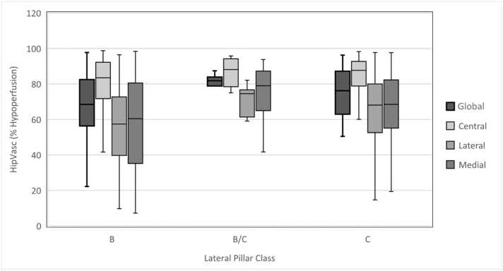
One-hundred and seven patients (88.8% male, median age 8.0 years) met the study criteria. The average global hypoperfusion was 73.7%. Poorer global perfusion was predictive of a longer duration of fragmentation (rho = 0.34, < .001) and of reossification (rho = 0.38, = .003). The average regional hypoperfusion of the medial, central, and lateral third of the femoral head was 65.3%, 83.7%, and 61.3% respectively, and was similarly related to the duration of fragmentation (rho = 0.26, 0.24, and 0.31, respectively) and of reossification (rho = 0.31, 0.43, and 0.39, respectively) ( < .05 for all). Similar to previous studies, we found a significant positive association between hypoperfusion in the lateral third of the femoral head and lateral pillar class ( = .037).
The degree of both global and regional hypoperfusion correlated with the duration of fragmentation and reossification stages in LCPD. Lateral epiphyseal hypoperfusion is predictive of lateral pillar class. Taken together, the information provided by perfusion magnetic resonance imaging can provide crucial prognostic information for children with LCPD.
1.Amount of hypoperfusion both globally and regionally on perfusion MRI correlates with the duration of fragmentation and reossification stages in LCPD.2.Lateral epiphyseal hypoperfusion correlates with lateral pillar class.3.Perfusion information offered by contrast MRI can offer crucial prognostic information in children with LCP.
II Prognostic Study.

Legg-Calvé-Perthes病(LCPD)通过4个具有独特影像学特征的阶段进展,阶段持续时间被认为是一个重要的预后因素。更新的灌注磁共振成像(pMRI)能够在疾病过程早期评估血管情况。本研究旨在描述早期pMRI上整体和局部灌注模式与Waldenström分期持续时间之间的关系。第二个目的是验证灌注不足与后续外侧柱分型之间的关系。
通过前瞻性收集的多中心国际队列,对早期LCPD(Waldenström I期)且行pMRI检查的患者每隔3个月进行系列X线片随访,至少随访2年。通过HipVasc软件对整个骨骺及股骨头的区域三等分量化骨骺灌注不足情况。Waldenström分期和外侧柱分型由3名小儿骨科医生通过模式评估确定。阶段持续时间定义为显示IIa期和IIIa期碎裂特征的第一张X线片之间以及IIIa期和IV期再骨化之间的间隔时间。
107例患者(88.8%为男性,中位年龄8.0岁)符合研究标准。整体平均灌注不足为73.7%。较差的整体灌注预示着碎裂期(rho = 0.34,P <.001)和再骨化期(rho = 0.38,P =.003)持续时间更长。股骨头内侧、中央和外侧三等分区域的平均局部灌注不足分别为65.3%、83.7%和61.3%,同样与碎裂期(rho分别为0.26、0.24和0.31)和再骨化期(rho分别为0.31、0.43和0.39)的持续时间相关(所有P <.05)。与既往研究相似,我们发现股骨头外侧三等分区域的灌注不足与外侧柱分型之间存在显著正相关(P =.037)。
整体和局部灌注不足程度均与LCPD的碎裂期和再骨化期持续时间相关。外侧骨骺灌注不足可预测外侧柱分型。综上所述,灌注磁共振成像提供的信息可为LCPD患儿提供关键的预后信息。
1.灌注MRI上整体和局部的灌注不足量与LCPD的碎裂期和再骨化期持续时间相关。2.外侧骨骺灌注不足与外侧柱分型相关。3.对比剂MRI提供的灌注信息可为LCP患儿提供关键的预后信息。
II级预后研究。