Qiu Jeffrey, Chen Min, Chen Zixin, Beinat Corinne, Melemenidis Stavros, Graves Edward, Rao Jianghong
Department of Radiology, Molecular Imaging Program at Stanford, Stanford University, Stanford, CA, 94305, USA.
Department of Radiation Oncology, Stanford University, Stanford, CA, 94305, USA.
EJNMMI Res. 2025 Jun 3;15(1):65. doi: 10.1186/s13550-025-01255-1.
Positron Emission Tomography (PET) imaging can monitor cancer treatment response by non-invasively detecting apoptosis in vivo. Signal-to-noise (SNR) remains one of the critical barriers to approval for clinical use. We have previously developed a PET tracer [18 F]-C-SNAT4 for imaging capase-3 activity in apoptotic tumors induced by chemo- and immunotherapy. [18 F]-C-SNAT4 is designed to undergo caspase-3 activated intramolecular cyclization. The product then self-assembles in situ into nanoparticles to generate preferential retention of F18 radioactivity in apoptotic cells. This unique mechanism prompted us to investigate if a cold mixture could enhance the probe retention and further augment the sensitivity for imaging radiotherapy.
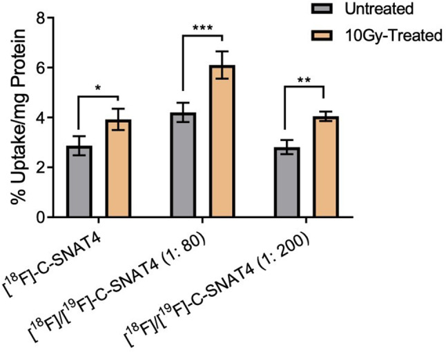
[18 F]-C-SNAT4 and hot/cold mixture [18 F]/[19 F]-C-SNAT4 were used to detect human NSCLC (NCI-H460) apoptosis induced by radiation. Both hot [18 F]-C-SNAT4 and hot/cold mixture [18 F]/[19 F]-C-SNAT4 had significantly increased uptake in radiation treated vs. untreated NCI-H460 cells in vitro. A 1: 80 hot/cold mixture increased signal by 1.6x compared to [18 F]-C-SNAT4 alone. In vivo studies were performed in murine xenograft models in high-dose radiation and low-dose radiation treatment groups. The hot/cold mixture showed an increase in the signal by 2.5x in high-dose radiation treated murine NCI-H460 xenograft models. Low-dose radiation induced apoptosis was only detected with the hot/cold mixture with 2.4x signal compared to hot [18 F]-C-SNAT4. Toxicity and dosimetry safety were evaluated at 250x and 10x respective dosages, then normalized to human dose equivalent.
A hot/cold mixture of [18 F]/[19 F]-C-SNAT4 generates significantly more signal compared to hot [18 F]-C-SNAT4, leading to higher sensitivity in detecting treatment response. This may present a solution to low sensitivity in the translation of apoptosis-specific radionuclides to clinical application.
正电子发射断层扫描(PET)成像可通过在体内非侵入性检测细胞凋亡来监测癌症治疗反应。信噪比(SNR)仍然是其获批临床应用的关键障碍之一。我们之前开发了一种PET示踪剂[18F]-C-SNAT4,用于对化疗和免疫疗法诱导的凋亡肿瘤中的半胱天冬酶-3活性进行成像。[18F]-C-SNAT4旨在经历半胱天冬酶-3激活的分子内环化。然后产物在原位自组装成纳米颗粒,以在凋亡细胞中产生F18放射性的优先保留。这种独特的机制促使我们研究冷混合物是否可以增强探针保留并进一步提高放射治疗成像的灵敏度。
使用[18F]-C-SNAT4和热/冷混合物[18F]/[19F]-C-SNAT4来检测辐射诱导的人非小细胞肺癌(NCI-H460)细胞凋亡。在体外,热的[18F]-C-SNAT4和热/冷混合物[18F]/[19F]-C-SNAT4在接受辐射治疗的NCI-H460细胞中的摄取均显著高于未接受治疗的细胞。与单独的[18F]-C-SNAT4相比,1:80的热/冷混合物使信号增加了1.6倍。在高剂量辐射和低剂量辐射治疗组的小鼠异种移植模型中进行了体内研究。在高剂量辐射治疗的小鼠NCI-H460异种移植模型中,热/冷混合物的信号增加了2.5倍。低剂量辐射诱导的细胞凋亡仅在热/冷混合物中被检测到,与热的[18F]-C-SNAT4相比,信号增加了2.4倍。分别在250倍和10倍剂量下评估了毒性和剂量测定安全性,然后将其归一化为人体等效剂量。
与热的[18F]-C-SNAT4相比,[18F]/[19F]-C-SNAT4的热/冷混合物产生的信号显著更多,从而在检测治疗反应方面具有更高的灵敏度。这可能为将凋亡特异性放射性核素转化为临床应用时灵敏度较低的问题提供一种解决方案。