Chen Charles D, Franklin Erin E, Li Yan, Joseph-Mathurin Nelly, Burns Aime L, Hobbs Diana A, McCullough Austin A, Schultz Stephanie A, Xiong Chengjie, Wang Guoqiao, Masellis Mario, Hsiung Ging-Yuek Robin, Gauthier Serge, Berman Sarah B, Roberson Erik D, Honig Lawrence S, Clarnette Roger, Ringman John M, Galvin James E, Brooks William, Suzuki Kazushi, Black Sandra, Levin Johannes, Aggarwal Neelum T, Jucker Mathias, Frosch Matthew P, Kofler Julia K, White Charles, Keene C Dirk, Chen Jie, Daniels Alisha, Gordon Brian A, Ibanez Laura, Karch Celeste M, Llibre-Guerra Jorge, McDade Eric, Morris John C, Supnet-Bell Charlene, Allegri Ricardo F, Lee Jae-Hong, Day Gregory S, Lopera Francisco, Roh Jee Hoon, Schofield Peter R, Mills Susan, Benzinger Tammie L S, Bateman Randall J, Perrin Richard J
Washington University in St. Louis, St. Louis, MO, USA.
Massachusetts General Hospital, Boston, MA, USA.
Acta Neuropathol. 2025 Jun 3;149(1):57. doi: 10.1007/s00401-025-02890-7.
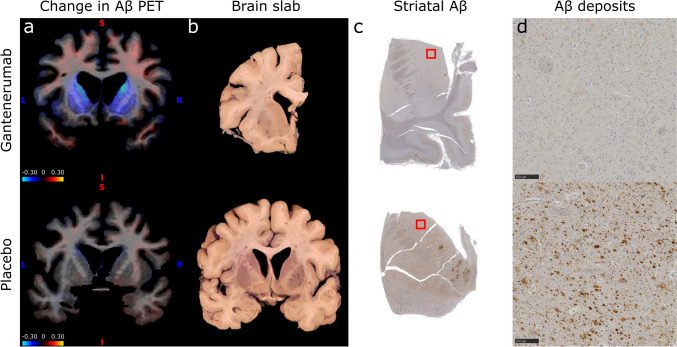
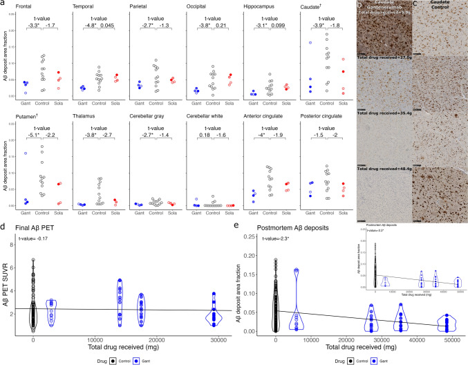
Clinical trials of anti-amyloid-β (Aβ) monoclonal antibodies in Alzheimer disease (AD) infer target engagement from Aβ positron emission tomography (PET) and/or fluid biomarkers such as cerebrospinal fluid (CSF) Aβ42/40. However, these biomarkers measure brain Aβ deposits indirectly and/or incompletely. In contrast, neuropathologic assessments allow direct investigation of treatment effects on brain Aβ deposits-and on potentially myriad 'downstream' pathologic features. From a clinical trial of anti-Aβ monoclonal antibodies in dominantly inherited AD (DIAD), in the largest study of its kind, we measured immunohistochemistry area fractions (AFs) for Aβ deposits (10D5), tauopathy (PHF1), microgliosis (IBA1), and astrocytosis (GFAP) in 10 brain regions from 10 trial cases-gantenerumab (n = 4), solanezumab (n = 4), placebo/no treatment (n = 2)-and 10 DIAD observational study cases. Strikingly, in proportion to total drug received, Aβ deposit AFs were significantly lower in the gantenerumab arm versus controls in almost all areas examined, including frontal, temporal, parietal, and occipital cortices, anterior cingulate, hippocampus, caudate, putamen, thalamus, and cerebellar gray matter; only posterior cingulate and cerebellar white matter comparisons were non-significant. In contrast, AFs of tauopathy, microgliosis, and astrocytosis showed no differences across groups. Our results demonstrate with direct histologic evidence that gantenerumab treatment in DIAD can reduce parenchymal Aβ deposits throughout the brain in a dose-dependent manner, suggesting that more complete removal may be possible with earlier and more aggressive treatment regimens. Although AFs of tauopathy, microgliosis, and astrocytosis showed no clear response to partial Aβ removal in this limited autopsy cohort, future examination of these cases with more sensitive techniques (e.g., mass spectrometry) may reveal more subtle 'downstream' effects.
抗淀粉样蛋白-β(Aβ)单克隆抗体治疗阿尔茨海默病(AD)的临床试验通过Aβ正电子发射断层扫描(PET)和/或脑脊液(CSF)Aβ42/40等液体生物标志物来推断靶点参与情况。然而,这些生物标志物只能间接和/或不完全地测量脑内Aβ沉积。相比之下,神经病理学评估能够直接研究治疗对脑内Aβ沉积以及潜在众多“下游”病理特征的影响。在一项针对显性遗传性AD(DIAD)的抗Aβ单克隆抗体临床试验中,在同类研究中规模最大的一项研究里,我们对10例试验病例(甘特单抗组n = 4、索拉单抗组n = 4、安慰剂/未治疗组n = 2)以及10例DIAD观察性研究病例的10个脑区进行了Aβ沉积(10D5)、tau蛋白病(PHF1)、小胶质细胞增生(IBA1)和星形细胞增生(GFAP)的免疫组化面积分数(AFs)测量。令人惊讶的是,与接受的总药物量成比例,在几乎所有检查区域,包括额叶、颞叶、顶叶和枕叶皮质、前扣带回、海马体、尾状核、壳核、丘脑和小脑灰质,甘特单抗组的Aβ沉积AFs显著低于对照组;只有后扣带回和小脑白质的比较无统计学意义。相比之下,tau蛋白病、小胶质细胞增生和星形细胞增生的AFs在各组之间没有差异。我们的结果通过直接的组织学证据表明,DIAD患者接受甘特单抗治疗可使全脑实质Aβ沉积以剂量依赖方式减少,这表明采用更早且更积极的治疗方案可能实现更完全的清除。尽管在这个有限的尸检队列中,tau蛋白病、小胶质细胞增生和星形细胞增生的AFs对部分Aβ清除没有明显反应,但未来使用更敏感技术(如质谱分析)对这些病例进行检查可能会揭示更细微的“下游”效应。