Dao Hien Thy, Loh Tiing Jen, Sharma Ravi K, Klareskog Lars, Malmström Vivianne, Reid Hugh H, Rossjohn Jamie, Lim Jia Jia
Infection and Immunity Program and Department of Biochemistry and Molecular Biology, Biomedicine Discovery Institute, Monash University, Clayton, Australia.
Division of Rheumatology, Department of Medicine, Karolinska Institutet, Karolinska University Hospital, Stockholm, Sweden; Center for Molecular Medicine, Karolinska Institutet, Solna, Sweden; Department of Clinical Immunology and Rheumatology, All India Institute of Medical Sciences, Bilaspur (H.P), India.
J Biol Chem. 2025 Jun 2;301(7):110326. doi: 10.1016/j.jbc.2025.110326.
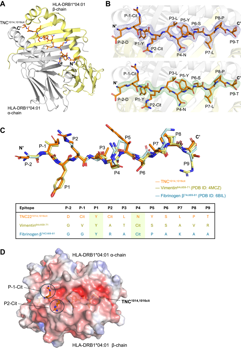
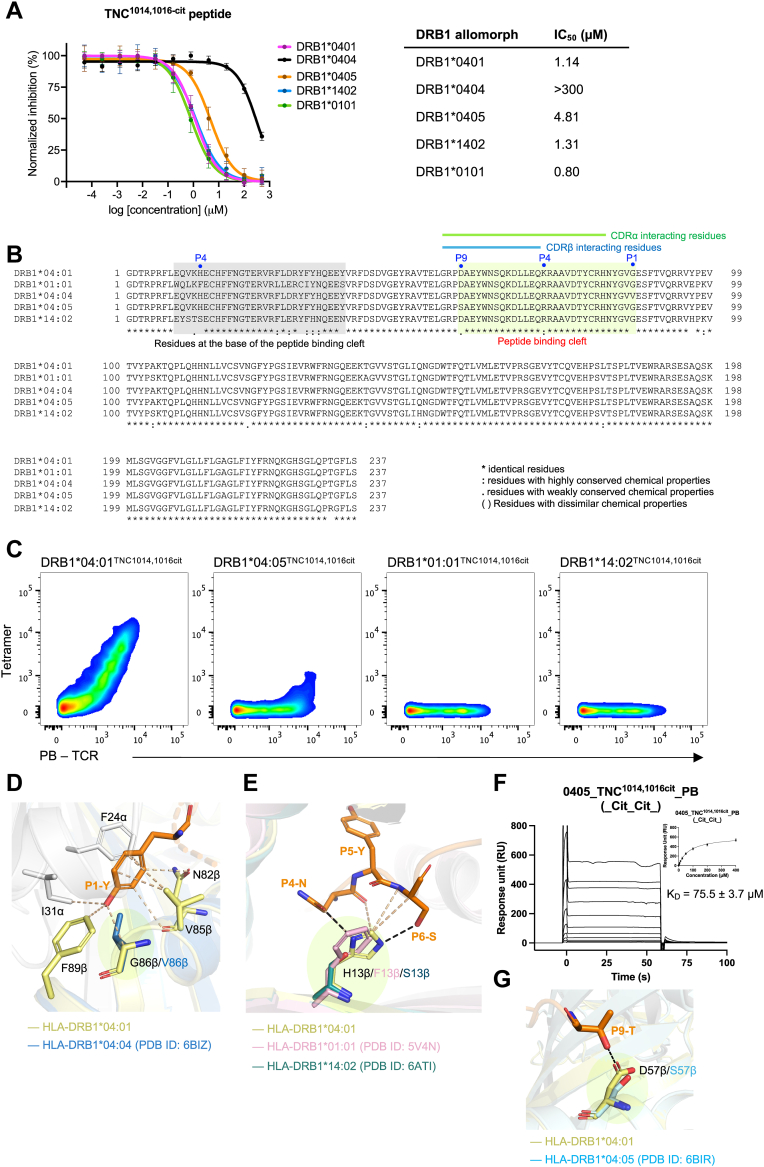
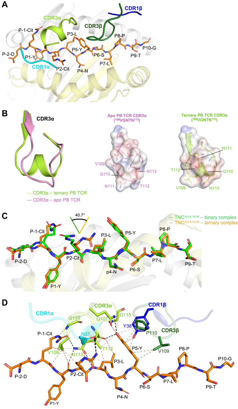
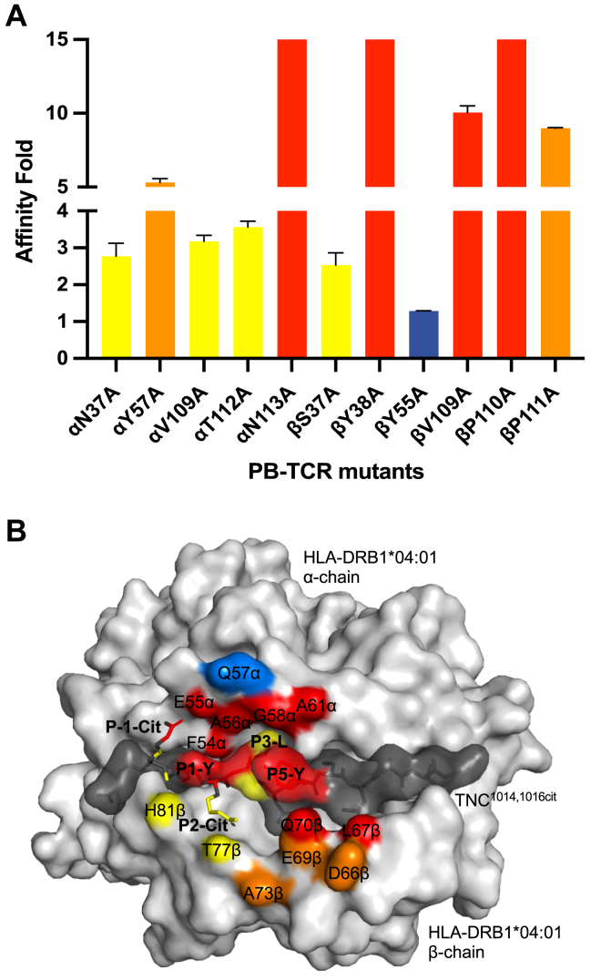
CD4 T cell autoreactivity against citrullinated (cit) self-epitopes presented by HLA-DRB1 is associated with rheumatoid arthritis (RA) pathogenesis. We understand the molecular bases of T cell receptor (TCR) recognition of cit-fibrinogen, cit-vimentin, and cit-α-enolase epitopes, and the role of citrulline in shaping TCR repertoire usage. Nevertheless, how TCRs recognize other cit-epitopes, including tenascin-C (TNC) and how alternative citrullination positions may modulate the T cell recognition remains unclear. Here, we examined TNC peptide, which contains citrullination at position P-1 and P2, to study the underlying TCR-HLA-DRB104:01-TNC molecular interactions. Crystal structure of HLA-DRB104:01 at 2.4 Å resolution revealed a conserved peptide binding register to the established HLA-DRB104:01-peptide structures, where both citrullines protruded upward. Next, we determined the crystal structure of a RA patient-derived TRAV35/TRBV10-2 (PB) TCR in complex with HLA-DRB104:01 at 3.2 Å resolution. The CDR3α loop (VGNTN) of PB TCR formed a secondary helical conformation at the N-terminus of the peptide binding cleft, allowing extensive interactions between the P-1 and P2 citrullines of TNC peptide. Surface plasmon resonance, tetramer staining, and CD69 activation assays revealed that the PB TCR did not cross-react to other RA autoantigens, and the P-1-Cit, P2-Cit, and P5-Tyr of TNC are the key determinants underlying the strict specificity of the PB TCR. Collectively, we provide molecular insight into citrullination in modulating TCR recognition.
针对由HLA - DRB1呈递的瓜氨酸化(cit)自身表位的CD4 T细胞自身反应性与类风湿性关节炎(RA)的发病机制相关。我们了解了T细胞受体(TCR)对瓜氨酸化纤维蛋白原、瓜氨酸化波形蛋白和瓜氨酸化α -烯醇化酶表位的识别分子基础,以及瓜氨酸在塑造TCR库使用中的作用。然而,TCR如何识别其他瓜氨酸化表位,包括肌腱蛋白 - C(TNC),以及不同的瓜氨酸化位置如何调节T细胞识别仍不清楚。在此,我们研究了在P - 1和P2位置含有瓜氨酸化的TNC肽,以探究潜在的TCR - HLA - DRB104:01 - TNC分子相互作用。分辨率为2.4 Å的HLA - DRB104:01晶体结构显示,其与已确定的HLA - DRB104:01 - 肽结构具有保守肽结合配准,其中两个瓜氨酸向上突出。接下来,我们以3.2 Å的分辨率确定了一名RA患者来源的TRAV35/TRBV10 - 2(PB)TCR与HLA - DRB104:01复合物的晶体结构。PB TCR的CDR3α环(VGNTN)在肽结合裂隙的N端形成二级螺旋构象,使得TNC肽的P - 1和P2瓜氨酸之间能够广泛相互作用。表面等离子体共振、四聚体染色和CD69激活试验表明,PB TCR不会与其他RA自身抗原发生交叉反应,并且TNC的P - 1 - Cit、P2 - Cit和P5 - Tyr是PB TCR严格特异性的关键决定因素。总体而言,我们提供了瓜氨酸化在调节TCR识别方面的分子见解。