Preziosa Paolo, Pagani Elisabetta, Margoni Monica, Rubin Martina, Storelli Loredana, Corazzolla Gianluca, Rocca Maria A, Filippi Massimo
Neuroimaging Research Unit, Division of Neuroscience, IRCCS San Raffaele Scientific Institute, Milan, Italy.
Neurology Unit, IRCCS San Raffaele Scientific Institute, Milan, Italy.
Neurol Neuroimmunol Neuroinflamm. 2025 Jul;12(4):e200414. doi: 10.1212/NXI.0000000000200414. Epub 2025 Jun 5.
The choroid plexus (CP) regulates immune functions and produces most CSF that circulates in the brain parenchyma through perivascular spaces, part of the glymphatic system. In multiple sclerosis (MS), CP enlargement and glymphatic dysfunction are associated with inflammatory activity, clinical disability, and brain damage, but their interrelation is unclear. We investigated whether glymphatic system dysfunction mediates the association between CP enlargement and brain damage in patients with MS.
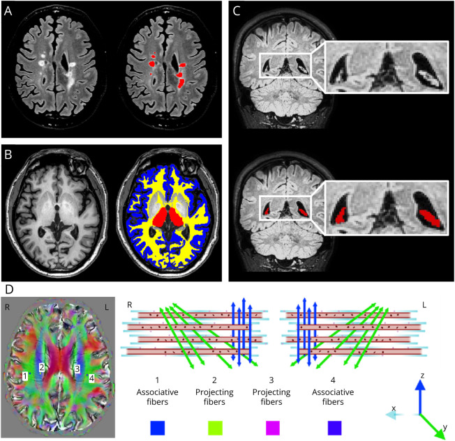
Brain fluid-attenuated inversion recovery, 3-dimensional T1-weighted, diffusion-weighted, and susceptibility-weighted sequences were obtained from 146 patients with MS and 72 healthy controls (HC). Glymphatic function was assessed using the diffusion along the perivascular space (DTI-ALPS) index, and CP volume was measured automatically.
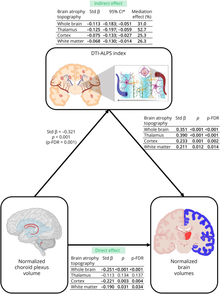
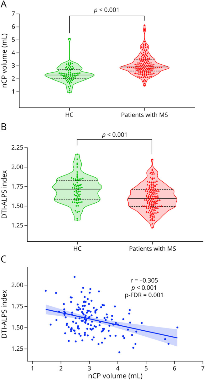
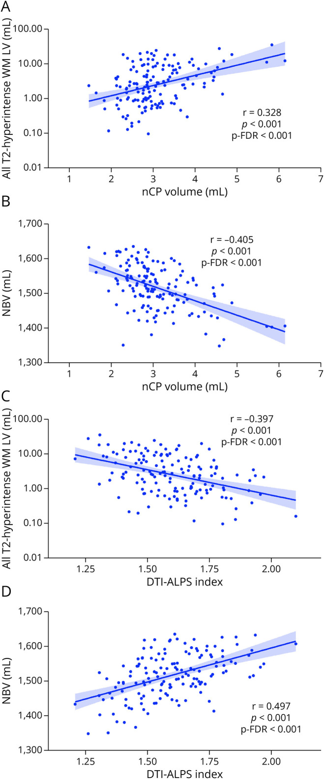
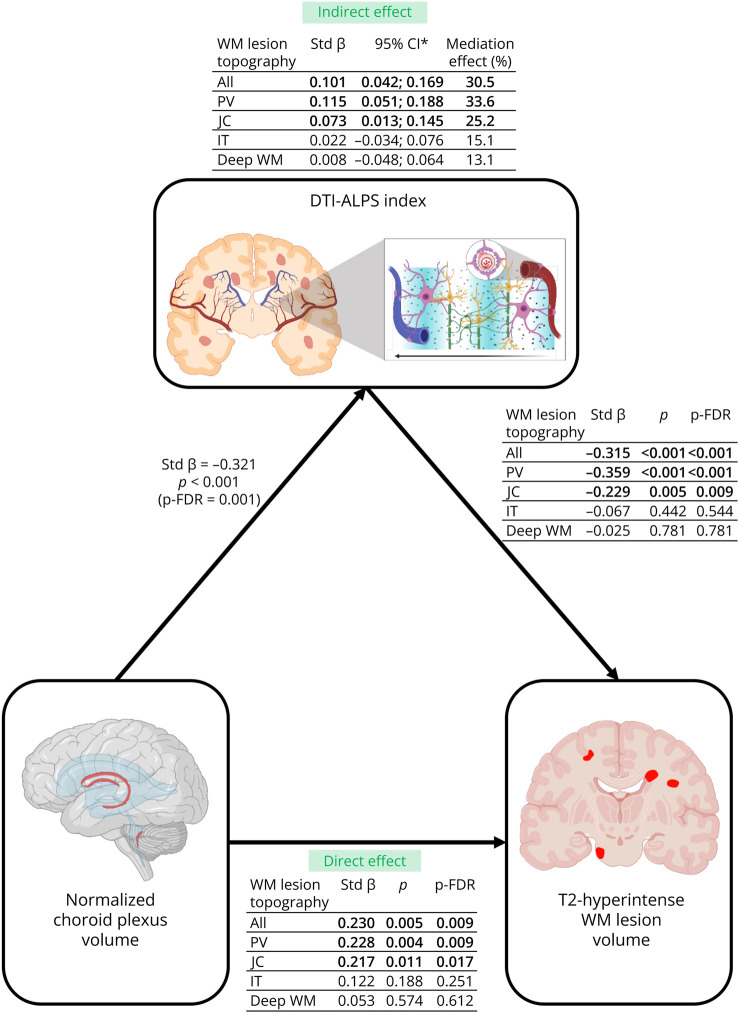
Patients with MS showed significantly higher white matter (WM) lesion and CP volumes ( < 0.001), and lower DTI-ALPS index, brain, WM, thalamic, and cortical volumes than HC ( ≤ 0.048). In patients with MS, higher CP volume correlated with a lower DTI-ALPS index ( = -0.305, false discovery rate p value = 0.001). Both measures were associated with higher total, periventricular, and juxtacortical (JC) WM lesion volumes (CP volume: from 0.285 to 0.340, p-FDR ≤ 0.001; DTI-ALPS index: from -0.301 to -0.444, ≤ 0.001), and lower brain, thalamic, cortical, and WM volumes (CP volume: from -0.246 to -0.405, p-FDR ≤ 0.006; DTI-ALPS index: from 0.269 to 0.497, p-FDR ≤ 0.003). The DTI-ALPS index partially mediated the associations of normalized choroid plexus volume with total, periventricular, and JC T2-hyperintense WM lesion volumes (standardized-β ranging from 0.073 to 0.115, relative effect ranging from 25.2% to 33.6%) and normalized brain, thalamic, cortical, and WM volumes (standardized-β ranging from -0.086 to -0.125, relative effect ranging from 25.3% to 52.7%).
In MS, enlarged normalized CP volume may contribute to brain damage accumulation possibly through the promotion of a chronic proinflammatory state and the mediation of glymphatic system dysfunction.
脉络丛(CP)调节免疫功能,并产生大部分通过血管周围间隙(淋巴系统的一部分)在脑实质中循环的脑脊液。在多发性硬化症(MS)中,脉络丛增大和淋巴系统功能障碍与炎症活动、临床残疾和脑损伤有关,但其相互关系尚不清楚。我们研究了淋巴系统功能障碍是否介导了MS患者脉络丛增大与脑损伤之间的关联。
对146例MS患者和72名健康对照者(HC)进行了脑液体衰减反转恢复序列、三维T1加权序列、扩散加权序列和磁敏感加权序列检查。使用沿血管周围间隙扩散(DTI-ALPS)指数评估淋巴系统功能,并自动测量脉络丛体积。
与HC相比,MS患者的白质(WM)病变和脉络丛体积显著更高(<0.001),而DTI-ALPS指数、脑、WM、丘脑和皮质体积更低(≤0.048)。在MS患者中,较高的脉络丛体积与较低的DTI-ALPS指数相关(=-0.305,错误发现率p值=0.001)。这两项指标均与更高的总脑室周围和皮质下(JC)WM病变体积相关(脉络丛体积:从0.285至0.340,p-FDR≤0.001;DTI-ALPS指数:从-0.301至-0.444,≤0.001),以及更低的脑、丘脑、皮质和WM体积(脉络丛体积:从-0.246至-0.405,p-FDR≤0.006;DTI-ALPS指数:从0.269至0.497,p-FDR≤0.003)。DTI-ALPS指数部分介导了标准化脉络丛体积与总脑室周围和JC T2高信号WM病变体积(标准化β范围为0.073至0.115,相对效应范围为25.2%至33.6%)以及标准化脑、丘脑、皮质和WM体积(标准化β范围为-0.086至-0.125,相对效应范围为25.3%至52.7%)之间的关联。
在MS中,标准化脉络丛体积增大可能通过促进慢性促炎状态和介导淋巴系统功能障碍,导致脑损伤积累。