Li Dongyu, Zhang Xinyu, Yang Fan, Jin Xin, Li Shumin, Yuan Haonan, Yao Lifen, Zhang Hong
Eye Hospital, The First Affiliated Hospital of Harbin Medical University, No.143, Yiman Street, Harbin City, Nangang District, Heilongjiang Province, China.
Department of Neurology, First Affiliated Hospital of Harbin Medical University, Harbin, Heilongjiang Province, China.
Invest Ophthalmol Vis Sci. 2025 Jun 2;66(6):21. doi: 10.1167/iovs.66.6.21.
To investigate whether the cornea nerve can distinguish between different subtypes of Parkinson's disease.
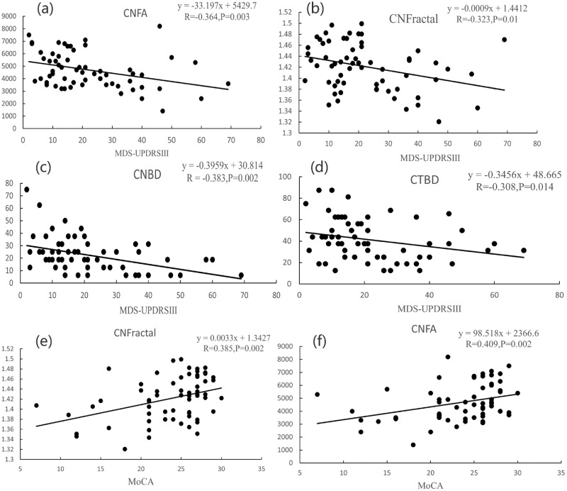
A total of 63 patients diagnosed with Parkinson's disease-comprising tremor-dominant (TD), postural instability and gait disturbance (PIGD), and mixed subtypes-were included alongside 31 age- and gender-matched control participants. All participants underwent In vivo confocal microscopy (IVCM) examinations along with comprehensive assessments of clinical neurological symptoms using the Movement Disorders Society Unified Parkinson's Disease Rating Scale, Hoehn and Yahr stages, and Montreal Cognitive Assessment scores. The detection range of IVCM includes the indicators of central and inferior whorl-like cornea nerve.
This study involved 63 patients, 23 were classified as having the TD type, 30 as having the PIGD type, and 10 as mixed type. Among them, most of central and whorl-like corneal nerve indicators were significantly lower in the PIGD group compared to the TD group. Receiver operating characteristic analysis demonstrated that combined central and inferior whorl-like corneal nerve indicators exhibited high discriminatory power between TD and PIGD types, with an area under the curve of 0.969.
As a non-invasive examination method, IVCM holds significant value for differentiating Parkinson's disease subtypes and identifying patients with varying motor manifestations. Among these findings, individuals with PIGD displayed more pronounced corneal nerve damage; furthermore, patients exhibiting lower inferior whorl length, corneal nerve fiber width, and fractal dimension of corneal nerves values were found to be at greater risk of being classified within the PIGD subtype.
研究角膜神经能否区分帕金森病的不同亚型。
纳入63例诊断为帕金森病的患者,包括震颤为主型(TD)、姿势不稳和步态障碍型(PIGD)以及混合型,同时纳入31名年龄和性别匹配的对照参与者。所有参与者均接受了共聚焦显微镜活体检查(IVCM),并使用运动障碍协会统一帕金森病评定量表、霍恩和雅尔分期以及蒙特利尔认知评估分数对临床神经症状进行了全面评估。IVCM的检测范围包括中央和下方螺旋状角膜神经的指标。
本研究纳入63例患者,其中23例为TD型,30例为PIGD型,10例为混合型。其中,与TD组相比,PIGD组的大多数中央和螺旋状角膜神经指标显著降低。受试者工作特征分析表明,中央和下方螺旋状角膜神经指标联合显示出对TD型和PIGD型的高鉴别力,曲线下面积为0.969。
作为一种非侵入性检查方法,IVCM在区分帕金森病亚型和识别具有不同运动表现的患者方面具有重要价值。在这些发现中,PIGD患者表现出更明显的角膜神经损伤;此外,发现下方螺旋长度、角膜神经纤维宽度和角膜神经分形维数较低的患者被归类为PIGD亚型的风险更大。