Muñoz Vanesa, Angulo-Ruiz Brenda Y, Gómez Carlos M
Human Psychobiology Laboratory, Experimental Psychology Department, University of Sevilla, Seville, Spain.
Cogn Neurodyn. 2025 Dec;19(1):88. doi: 10.1007/s11571-025-10281-7. Epub 2025 Jun 9.
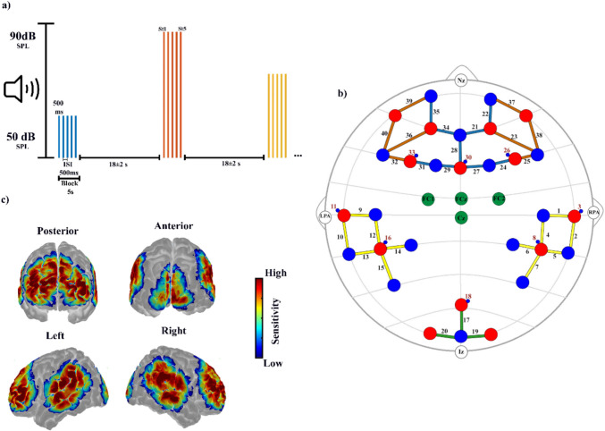
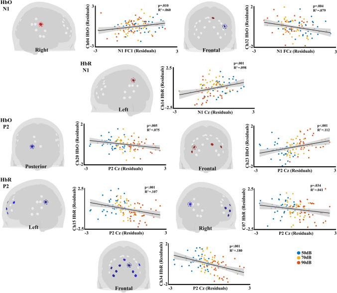
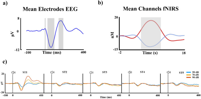
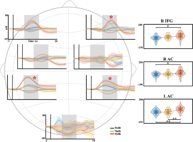
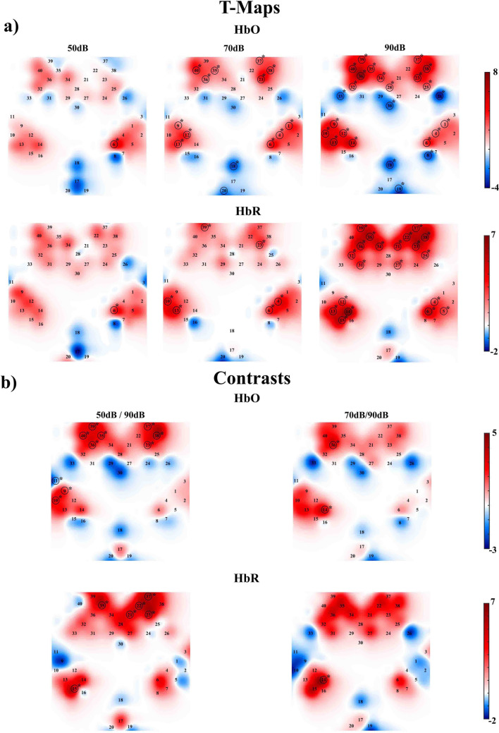
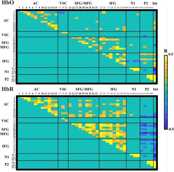
Recent studies combining electroencephalography (EEG) and functional near-infrared spectroscopy (fNIRS) have shown promising results linking neural and vascular responses. This study analyzes the topographical effect of auditory stimulus intensity on cortical activation and explores neurovascular coupling between fNIRS hemodynamic signals and auditory-evoked potentials (AEPs), extracted from EEG. Forty healthy volunteers (13 males, 27 females; mean age = 22.27 ± 3.96 years) listened to complex tones of varying intensities (50-, 70-, and 90-dB SPL) across seven frequencies (range of 400-2750 Hz) in blocks of five, while EEG and fNIRS were recorded. PERMANOVA analysis revealed that increasing intensity modulated hemodynamic activity, leading to amplitude changes and enhanced recruitment of auditory and prefrontal cortices. To isolate stimulus-specific activity, Spearman correlations were computed on residuals-components of AEPs and fNIRS responses with individual trends removed. The N1 amplitude increase was correlated with higher superior temporal gyrus (STG) and superior frontal gyrus (SFG) activity, and reduced activity in inferior frontal gyrus (IFG) for the oxygenated hemoglobin (HbO), while the deoxygenated hemoglobin (HbR) was associated with increased activity in one channel near the Supramarginal Gyrus (SMG). P2 amplitude increase was associated with higher activation in SFG and IFG for HbO, while for HbR with the activity in SMG, angular gyrus (AnG), SFG, and IFG. Additionally, internal correlations between fNIRS channels revealed strong associations within auditory and frontal regions. These findings provide insights into existing models of neurovascular coupling by showing how stimulus properties, such as intensity, modulate the relationship between neural activity and vascular responses.
The online version contains supplementary material available at 10.1007/s11571-025-10281-7.
最近将脑电图(EEG)和功能近红外光谱(fNIRS)相结合的研究显示了将神经反应与血管反应联系起来的有前景的结果。本研究分析了听觉刺激强度对皮层激活的地形学效应,并探索了从EEG中提取的fNIRS血流动力学信号与听觉诱发电位(AEP)之间的神经血管耦合。40名健康志愿者(13名男性,27名女性;平均年龄 = 22.27 ± 3.96岁)以五个为一组,聆听了七个频率(400 - 2750 Hz范围)下不同强度(50、70和90 dB SPL)的复杂音调,同时记录EEG和fNIRS。PERMANOVA分析显示,强度增加调节了血流动力学活动,导致幅度变化并增强了听觉和前额叶皮层的募集。为了分离特定刺激活动,对去除个体趋势后的AEP和fNIRS反应的残差成分计算了斯皮尔曼相关性。N1波幅增加与颞上回(STG)和额上回(SFG)更高的活动相关,而对于氧合血红蛋白(HbO),额下回(IFG)的活动减少,而脱氧血红蛋白(HbR)与缘上回(SMG)附近一个通道的活动增加相关。P2波幅增加与HbO的SFG和IFG更高的激活相关,而对于HbR与SMG、角回(AnG)、SFG和IFG的活动相关。此外,fNIRS通道之间的内部相关性显示听觉和额叶区域内有很强的关联。这些发现通过展示刺激特性(如强度)如何调节神经活动与血管反应之间的关系,为现有的神经血管耦合模型提供了见解。
在线版本包含可在10.1007/s11571-025-10281-7获取的补充材料。