Santiago Joana, Silva Joana V, Santos Manuel A S, Fardilha Margarida
Department of Medical Sciences, Institute of Biomedicine, University of Aveiro, 3810-193 Aveiro, Portugal.
Multidisciplinary Institute of Ageing, MIA-Portugal, University of Coimbra, 3000-370 Coimbra, Portugal.
Cells. 2025 May 30;14(11):813. doi: 10.3390/cells14110813.
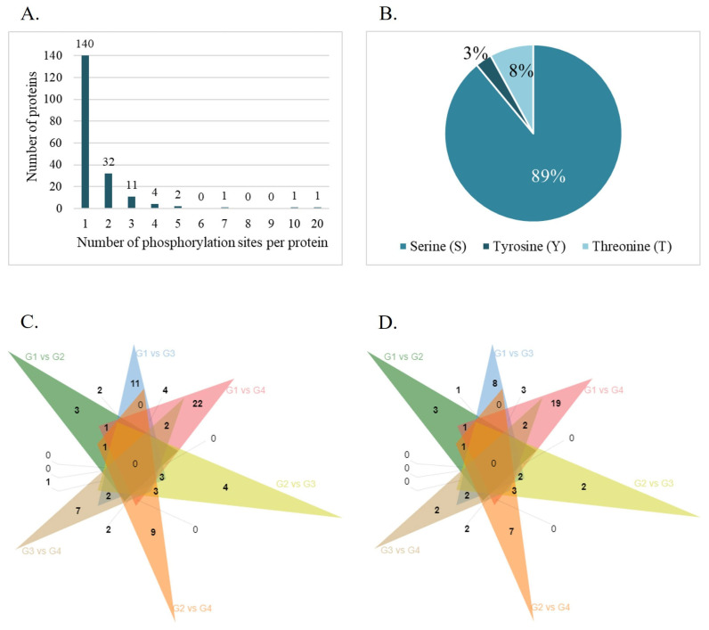
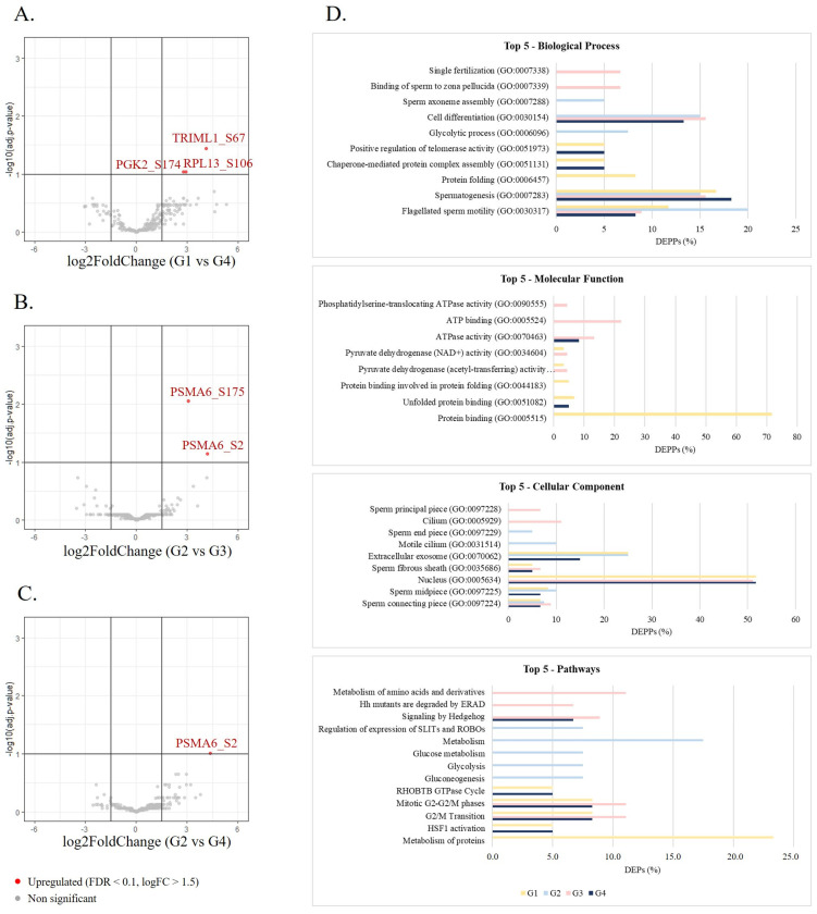
Male fertility is strongly influenced by environmental exposures, lifestyle, and advancing age. While advanced paternal age (APA) has been linked with a progressive decline in male fertility, poor reproductive outcomes, and decreased offspring health, the molecular mechanisms underlying these alterations remain unclear. In this work, we investigated the impact of men's age on human sperm protein expression and phosphorylation to identify molecular alterations possibly responsible for the age-associated decline in male fertility. Semen samples from volunteers attending fertility consultations at the Hospital of Aveiro were collected, analyzed according to WHO's guidelines, and processed by the density gradient technique. The proteome and phosphoproteome of 19 normozoospermic human sperm samples divided into four age groups were evaluated by mass spectrometry: ≤30 years old; 31-35 years old; 36-40 years old; and >40 years old. Proteomic analysis revealed 46 differentially expressed proteins (DEPs) between groups, some of them associated with infertility-related phenotypes. Gene ontology (GO) analysis, performed using the DAVID database, revealed that DEPs in older men were enriched in pathways related to stress response, metabolism, and embryo implantation. Additionally, 94 differentially phosphorylated sites corresponding to 76 differentially expressed phosphorylated proteins between the groups were identified, related to key reproductive processes such as sperm motility, spermatogenesis, and sperm binding to zona pellucida, and involved in metabolic and stress response pathways, like HSF1 activation. The set of proteins and phosphorylated residues altered in the sperm fraction usually used in assisted reproductive technology (ART) highlights the need to consider the age of the male partner during fertility assessment and treatment planning. These markers can also be used to explain cases of idiopathic infertility, failure in ART, or repeated abortion associated with APA, overcoming the subjectivity of the conventional semen analysis.
男性生育能力受到环境暴露、生活方式和年龄增长的强烈影响。虽然父亲年龄偏大(APA)与男性生育能力的逐渐下降、不良生殖结局以及后代健康状况下降有关,但这些改变背后的分子机制仍不清楚。在这项研究中,我们调查了男性年龄对人类精子蛋白质表达和磷酸化的影响,以确定可能导致与年龄相关的男性生育能力下降的分子改变。收集了在阿威罗医院参加生育咨询的志愿者的精液样本,按照世界卫生组织的指南进行分析,并采用密度梯度技术进行处理。通过质谱法评估了19份正常精子的人类精子样本的蛋白质组和磷酸化蛋白质组,这些样本分为四个年龄组:≤30岁;31 - 35岁;36 - 40岁;以及>40岁。蛋白质组学分析揭示了不同组之间有46种差异表达蛋白(DEP),其中一些与不育相关表型有关。使用DAVID数据库进行的基因本体(GO)分析表明,老年男性中的DEP在与应激反应、代谢和胚胎着床相关的途径中富集。此外,还鉴定出了94个差异磷酸化位点,对应于不同组之间76种差异表达的磷酸化蛋白,这些位点与精子活力、精子发生以及精子与透明带结合等关键生殖过程有关,并参与代谢和应激反应途径,如热休克因子1(HSF1)的激活。在辅助生殖技术(ART)中通常使用的精子部分中发生改变的蛋白质和磷酸化残基组突出表明在生育评估和治疗计划过程中需要考虑男性伴侣的年龄。这些标志物还可用于解释与APA相关的特发性不育、ART失败或反复流产的病例,克服了传统精液分析的主观性。