Zeng Gang, Sun Yujun, Liang Min, Liu Wenzhou, Chen Yanbo, Wu Jionglin, Zheng Jiayuan, Zhou Taolue, Zhao Huiying, Song Weidong, Ding Yue
Department of Orthopedics, Sun Yat-Sen Memorial Hospital, 107 Yanjiang West Road, Yuexiu District, Guangzhou, Guangdong Province, China.
Operating Theatre, Sun Yat-Sen Memorial Hospital, Guangzhou, Guangdong Province, China.
Eur J Med Res. 2025 Jun 14;30(1):480. doi: 10.1186/s40001-025-02687-0.
Talus is a critical component of the ankle joint that allows multidirectional movement essential for gait and mobility. Talus prostheses are typically used in cases of severe trauma, avascular necrosis, or end-stage arthritis. Adjacent joint arthritis is the most common complication after total talar replacement (TTR). While intervention is rarely required, varying degrees of postoperative degenerative changes have been reported in surrounding joints. Therefore, a detail evaluation of /the mechanical stability and stress distribution within talus prostheses is crucial. Recent advancements, including patient-specific implants and improved biomaterials, show promise in enhancing long-term outcomes and reducing complication rates.
This study investigates the mechanical stability and stress distribution changes in a talus prosthesis after the sequential removal of surrounding ligaments and the application of varying thicknesses of cartilage overlays.
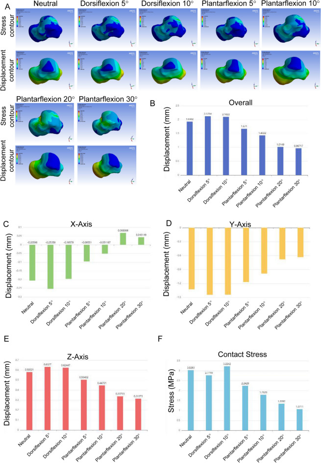
Finite element analysis (FEA) was employed using CT data to create a 3D model of the talus prosthesis. The model was refined using Mimics, Geomagic, and SolidWorks, and then analyzed with Ansys. The mechanical properties of bone, prosthesis, skin, ligament, and cartilage were incorporated into the model. Various ankle positions, including neutral, dorsiflexion (5° and 10°), and plantarflexion (5°, 10°, 15°, 20°and 30°), were simulated under different ligament removal conditions.
The sequential removal of surrounding ligaments, including lateral, medial, sinus tarsi, and talonavicular ligaments, did not significantly affect the stability of the talus prosthesis. Stress values ranged from 2.2257 to 3.0218 MPa, and displacement values were stable around 1.6611 to 2.2119 mm across different conditions. By comparing the effects of different cartilage thicknesses on contact area and contact stress across various flexion angles, we have the optimized the cartilage thickness, and identified a 0.5mm cartilage overlay resulted in stress distributions most similar to a normal ankle joint, enhancing the prosthesis performance.
The study demonstrates that the removal of surrounding ligaments does not compromise the stability of the talus prosthesis. Importantly, a 0.5 mm cartilage overlay is optimal for achieving stress distributions comparable to a normal ankle joint. These findings offer valuable preliminary insights into stress distribution patterns following talus prosthesis implantation; however, they represent an initial step, and further experimental and clinical studies are needed to validate and extend these results.
Level V, computational simulation study.
距骨是踝关节的关键组成部分,它允许进行对步态和活动能力至关重要的多方向运动。距骨假体通常用于严重创伤、缺血性坏死或终末期关节炎的病例。相邻关节关节炎是全距骨置换(TTR)后最常见的并发症。虽然很少需要干预,但已有报道称周围关节出现了不同程度的术后退行性改变。因此,详细评估距骨假体内部的机械稳定性和应力分布至关重要。包括定制植入物和改良生物材料在内的最新进展,在改善长期疗效和降低并发症发生率方面显示出前景。
本研究调查了在依次去除周围韧带并应用不同厚度的软骨覆盖物后距骨假体的机械稳定性和应力分布变化。
利用CT数据采用有限元分析(FEA)创建距骨假体的三维模型。使用Mimics、Geomagic和SolidWorks对模型进行优化,然后用Ansys进行分析。将骨骼、假体、皮肤、韧带和软骨的力学性能纳入模型。在不同的韧带去除条件下模拟了包括中立位、背屈(5°和10°)和跖屈(5°、10°、15°、20°和30°)在内的各种踝关节位置。
依次去除包括外侧、内侧、跗骨窦和距舟韧带在内的周围韧带,对距骨假体的稳定性没有显著影响。在不同条件下,应力值范围为2.2257至3.0218MPa,位移值稳定在1.6611至2.2119mm左右。通过比较不同软骨厚度对不同屈曲角度下接触面积和接触应力的影响,我们优化了软骨厚度,并确定0.5mm的软骨覆盖物导致的应力分布与正常踝关节最相似,从而提高了假体性能。
该研究表明去除周围韧带不会损害距骨假体的稳定性。重要的是,0.5mm的软骨覆盖物对于实现与正常踝关节相当的应力分布是最佳的。这些发现为距骨假体植入后的应力分布模式提供了有价值的初步见解;然而,它们只是初步步骤,需要进一步的实验和临床研究来验证和扩展这些结果。
V级,计算模拟研究。