• 文献检索
  • 文档翻译
  • 深度研究
  • 学术资讯
  • Suppr Zotero 插件Zotero 插件
  • 邀请有礼
  • 套餐&价格
  • 历史记录
应用&插件
Suppr Zotero 插件Zotero 插件浏览器插件Mac 客户端Windows 客户端微信小程序
定价
高级版会员购买积分包购买API积分包
服务
文献检索文档翻译深度研究API 文档MCP 服务
关于我们
关于 Suppr公司介绍联系我们用户协议隐私条款
关注我们

Suppr 超能文献

核心技术专利:CN118964589B侵权必究
粤ICP备2023148730 号-1Suppr @ 2026

文献检索

告别复杂PubMed语法,用中文像聊天一样搜索,搜遍4000万医学文献。AI智能推荐,让科研检索更轻松。

立即免费搜索

文件翻译

保留排版,准确专业,支持PDF/Word/PPT等文件格式,支持 12+语言互译。

免费翻译文档

深度研究

AI帮你快速写综述,25分钟生成高质量综述,智能提取关键信息,辅助科研写作。

立即免费体验

人类肥厚性室间隔的生物力学特性因心肌细胞增大、胶原蛋白过度表达和微观结构紊乱而恶化。

Deteriorated biomechanical properties of human hypertrophied septum in response to cardiomyocyte enlargement, overexpressed collagen, and disarrayed microstructures.

作者信息

Copeland Katherine M, Chen Houjia, Chintapula Uday, Almasian Milad, Chung Duc Khang, Taylor Alan M, Ding Yichen, Sharma Gaurav, Jessen Michael E, Hong Yi, Nguyen Kytai T, Peltz Matthias, Bajona Pietro, Liao Jun

机构信息

Department of Bioengineering, University of Texas at Arlington, Arlington, TX, United States.

Department of Bioengineering, University of Texas at Dallas, Richardson, TX, United States.

出版信息

Front Bioeng Biotechnol. 2025 Jun 2;13:1620594. doi: 10.3389/fbioe.2025.1620594. eCollection 2025.

DOI:10.3389/fbioe.2025.1620594
PMID:40529169
原文链接:https://pmc.ncbi.nlm.nih.gov/articles/PMC12171370/
Abstract

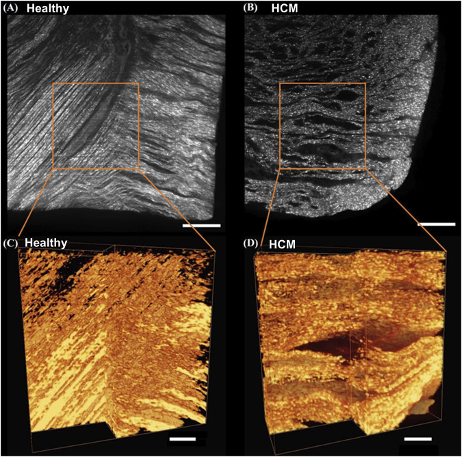
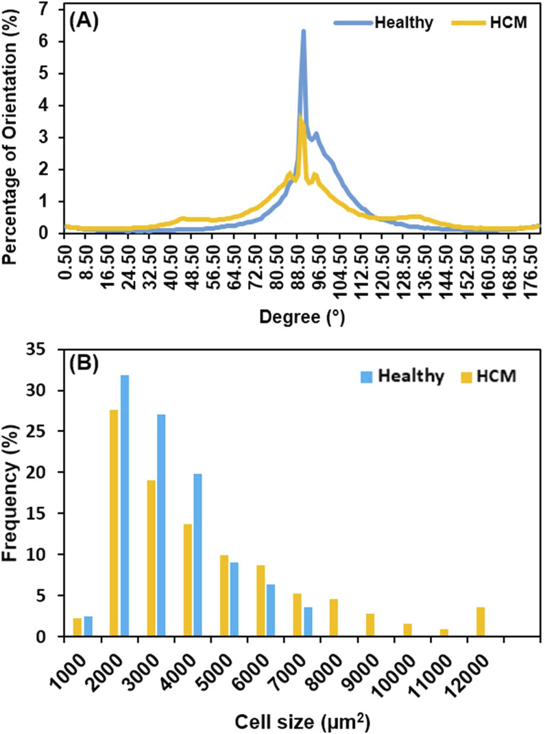
Hypertrophic cardiomyopathy (HCM) is often caused by genetic mutations, resulting in abnormal thickening of ventricular muscle, particularly the septum, and causing left ventricular outflow tract (LVOT) obstruction and inferior cardiac performance. The cell and microstructural abnormalities are believed to be the cause of the altered tissue mechanical properties and inferior performance. However, there is a lack of detailed biomechanical assessments of human hypertrophied septum and a lack of understanding of the structural-mechanical relationship between altered biomechanical properties and cellular hypertrophy, fibrotic overexpression, and microstructural disruptions. In this study, we performed thorough biomechanical and microstructural characterizations on the human hypertrophied septum and compared this with healthy septum. We found that the hypertrophied human septum was stiffer at the initial phase of tissue loading, but less nonlinear, less stiff in the linear region, and much weaker in mechanical strength when compared to the healthy human septum. The fibrosis-induced initial stiffening in the hypertrophied septum paradoxically coexists with compromised mechanical strength and integrity under physiological demands, correlating with the clinical observations of diastolic dysfunction and susceptibility to myocardial damage in HCM patients despite ventricular wall thickening. We also discovered that the human hypertrophied septum had significantly larger stress relaxation and slightly larger creep when compared to healthy septum. Moreover, the abnormal, disorganized cell-collagen microstructures in the hypertrophied septum make short-term stress release more difficult and require longer relaxation times to reach equilibrium. Biaxial testing performed at the initial phase of tissue loading showed that both the healthy septum and hypertrophied septum had nonlinear anisotropic stress-strain behavior and confirmed that, in the longitudinal direction, the hypertrophied septum was stiffer than the healthy septum. Our microstructural quantifications via histology and light-sheet microscopy revealed that (i) the heterogeneous cardiomyocyte enlargement and disarray, combined with disorganized collagen overexpression, create a mechanically inefficient tissue architecture in the hypertrophied septum, and (ii) the observed cell-collagen microstructural disruptions provide mechanistic explanations for the deteriorated biomechanical properties. Our viscoelastic mechanical data and microstructural characterizations build a strong foundation to understand the altered tissue behavior of the hypertrophied septum, the degree of deviation from the normal septum, and the underlying structural mechanisms.

摘要

肥厚型心肌病(HCM)通常由基因突变引起,导致心室肌异常增厚,尤其是室间隔增厚,并引起左心室流出道(LVOT)梗阻和心脏功能下降。细胞和微观结构异常被认为是组织力学性能改变和功能下降的原因。然而,目前缺乏对人类肥厚室间隔的详细生物力学评估,也缺乏对生物力学性能改变与细胞肥大、纤维化过度表达和微观结构破坏之间结构 - 力学关系的理解。在本研究中,我们对人类肥厚室间隔进行了全面的生物力学和微观结构表征,并与健康室间隔进行了比较。我们发现,与健康人类室间隔相比,肥厚的人类室间隔在组织加载的初始阶段更硬,但非线性程度较低,在线性区域更软,机械强度更弱。肥厚室间隔中由纤维化引起的初始硬化与生理需求下机械强度和完整性受损并存,这与HCM患者尽管心室壁增厚但仍出现舒张功能障碍和心肌损伤易感性的临床观察结果相关。我们还发现,与健康室间隔相比,人类肥厚室间隔的应力松弛明显更大,蠕变略大。此外,肥厚室间隔中异常、无序的细胞 - 胶原微观结构使短期应力释放更加困难,需要更长的松弛时间才能达到平衡。在组织加载初始阶段进行的双轴测试表明,健康室间隔和肥厚室间隔均具有非线性各向异性应力 -应变行为,并证实,在纵向方向上,肥厚室间隔比健康室间隔更硬。我们通过组织学和光片显微镜进行的微观结构定量分析表明:(i)心肌细胞的异质性增大和排列紊乱,加上无序的胶原过度表达,在肥厚室间隔中形成了机械效率低下的组织结构;(ii)观察到的细胞 - 胶原微观结构破坏为生物力学性能恶化提供了机制解释。我们的粘弹性力学数据和微观结构表征为理解肥厚室间隔的组织行为改变、与正常室间隔的偏离程度以及潜在的结构机制奠定了坚实基础。

https://cdn.ncbi.nlm.nih.gov/pmc/blobs/63dc/12171370/8135defa7766/fbioe-13-1620594-g006.jpg
https://cdn.ncbi.nlm.nih.gov/pmc/blobs/63dc/12171370/bc4b2a2e9db5/fbioe-13-1620594-g001.jpg
https://cdn.ncbi.nlm.nih.gov/pmc/blobs/63dc/12171370/ecea470c77fc/fbioe-13-1620594-g002.jpg
https://cdn.ncbi.nlm.nih.gov/pmc/blobs/63dc/12171370/5f3e6d4bc3c5/fbioe-13-1620594-g003.jpg
https://cdn.ncbi.nlm.nih.gov/pmc/blobs/63dc/12171370/2a501e7efd5d/fbioe-13-1620594-g004.jpg
https://cdn.ncbi.nlm.nih.gov/pmc/blobs/63dc/12171370/cbe35d0eeb53/fbioe-13-1620594-g005.jpg
https://cdn.ncbi.nlm.nih.gov/pmc/blobs/63dc/12171370/8135defa7766/fbioe-13-1620594-g006.jpg

相似文献

1
Deteriorated biomechanical properties of human hypertrophied septum in response to cardiomyocyte enlargement, overexpressed collagen, and disarrayed microstructures.人类肥厚性室间隔的生物力学特性因心肌细胞增大、胶原蛋白过度表达和微观结构紊乱而恶化。
Front Bioeng Biotechnol. 2025 Jun 2;13:1620594. doi: 10.3389/fbioe.2025.1620594. eCollection 2025.
2
Native and decellularized porcine vena cava: Biomechanical and microstructural comparison.天然及去细胞猪腔静脉:生物力学与微观结构比较。
Acta Biomater. 2025 May 31. doi: 10.1016/j.actbio.2025.05.071.
3
Adapting Safety Plans for Autistic Adults with Involvement from the Autism Community.在自闭症群体的参与下为成年自闭症患者调整安全计划。
Autism Adulthood. 2025 May 28;7(3):293-302. doi: 10.1089/aut.2023.0124. eCollection 2025 Jun.
4
Electronic cigarettes for smoking cessation.用于戒烟的电子烟。
Cochrane Database Syst Rev. 2025 Jan 29;1(1):CD010216. doi: 10.1002/14651858.CD010216.pub9.
5
Interventions for fertility preservation in women with cancer undergoing chemotherapy.对接受化疗的癌症女性进行生育力保存的干预措施。
Cochrane Database Syst Rev. 2025 Jun 19;6:CD012891. doi: 10.1002/14651858.CD012891.pub2.
6
Aural toilet (ear cleaning) for chronic suppurative otitis media.慢性化脓性中耳炎的耳道清理(耳部清洁)
Cochrane Database Syst Rev. 2025 Jun 9;6(6):CD013057. doi: 10.1002/14651858.CD013057.pub3.
7
An Occupational Science Contribution to Camouflaging Scholarship: Centering Intersectional Experiences of Occupational Disruptions.职业科学对伪装学术的贡献:以职业中断的交叉经历为中心
Autism Adulthood. 2025 May 28;7(3):238-248. doi: 10.1089/aut.2023.0070. eCollection 2025 Jun.
8
Non-pharmacological interventions for sleep promotion in hospitalized children.促进住院儿童睡眠的非药物干预措施。
Cochrane Database Syst Rev. 2022 Jun 15;6(6):CD012908. doi: 10.1002/14651858.CD012908.pub2.
9
Molecular feature-based classification of retroperitoneal liposarcoma: a prospective cohort study.基于分子特征的腹膜后脂肪肉瘤分类:一项前瞻性队列研究。
Elife. 2025 May 23;14:RP100887. doi: 10.7554/eLife.100887.
10
Absorbent products for moderate-heavy urinary and/or faecal incontinence in women and men.适用于女性和男性中重度尿失禁和/或大便失禁的吸收性产品。
Cochrane Database Syst Rev. 2008 Oct 8(4):CD007408. doi: 10.1002/14651858.CD007408.

本文引用的文献

1
Mavacamten (CAMZYOS) First-in-Class Small-Molecule Myosin Inhibitor for Treatment of Obstructive Hypertrophic Cardiomyopathy.马伐卡坦(CAMZYOS):用于治疗梗阻性肥厚型心肌病的首款小分子肌球蛋白抑制剂。
Clin Nurse Spec. 2024;38(3):119-121. doi: 10.1097/NUR.0000000000000821.
2
Light-Sheet Imaging to Reveal Cardiac Structure in Rodent Hearts.光片成像揭示啮齿动物心脏的心脏结构
J Vis Exp. 2024 Mar 29(205). doi: 10.3791/66707.
3
Mavacamten: a first-in-class myosin inhibitor for obstructive hypertrophic cardiomyopathy.马卡丹特:一种用于梗阻性肥厚型心肌病的首创肌球蛋白抑制剂。
Eur Heart J. 2023 Nov 21;44(44):4622-4633. doi: 10.1093/eurheartj/ehad637.
4
Light sheet imaging and interactive analysis of the cardiac structure in neonatal mice.光片成像和新生鼠心脏结构的交互式分析。
J Biophotonics. 2023 May;16(5):e202200278. doi: 10.1002/jbio.202200278. Epub 2023 Jan 18.
5
Strain-dependent stress relaxation behavior of healthy right ventricular free wall.健康右心室游离壁的应变依赖性应力松弛行为。
Acta Biomater. 2022 Oct 15;152:290-299. doi: 10.1016/j.actbio.2022.08.043. Epub 2022 Aug 24.
6
Biomechanical properties of acellular scar ECM during the acute to chronic stages of myocardial infarction.心肌梗死急性至慢性阶段无细胞瘢痕细胞外基质的生物力学特性。
J Mech Behav Biomed Mater. 2021 Apr;116:104342. doi: 10.1016/j.jmbbm.2021.104342. Epub 2021 Jan 22.
7
Collagen Cross-Linking Is Associated With Cardiac Remodeling in Hypertrophic Obstructive Cardiomyopathy.胶原交联与肥厚型梗阻性心肌病的心脏重构相关。
J Am Heart Assoc. 2021 Jan 5;10(1):e017752. doi: 10.1161/JAHA.120.017752. Epub 2020 Dec 25.
8
Epicardial prestrained confinement and residual stresses: a newly observed heart ventricle confinement interface.心外膜预拉伸约束和残余应力:一种新观察到的心脏心室约束界面。
J R Soc Interface. 2019 Mar 29;16(152):20190028. doi: 10.1098/rsif.2019.0028.
9
Decreased biventricular mechanics and functional reserve in nonobstructive hypertrophic cardiomyopathy patients: implications for exercise capacity.非梗阻性肥厚型心肌病患者双心室力学及功能储备降低:对运动能力的影响
Int J Cardiovasc Imaging. 2019 May;35(5):869-879. doi: 10.1007/s10554-019-01530-y. Epub 2019 Jan 17.
10
Light-sheet Fluorescence Microscopy for the Study of the Murine Heart.用于研究小鼠心脏的光片荧光显微镜技术
J Vis Exp. 2018 Sep 15(139):57769. doi: 10.3791/57769.