Feng Shi, Yang Jingyuan, Zhao Xinyu, Zhao Jianchun, Du Yunfeng, Yu Weihong, Ding Dayong, Li Xirong, Chen Youxin
Ophthalmology Department, Peking Union Medical College Hospital, Beijing, China.
Vistel AI Lab, Visionary Intelligence Ltd, Beijing, China.
Front Cell Dev Biol. 2025 Jun 4;13:1609567. doi: 10.3389/fcell.2025.1609567. eCollection 2025.
The aim of this study is to generate post-therapeutic optical coherence tomography (OCT) images based on pre-therapeutic OCT by using generative adversarial networks (GANs). The synthetic images enable us to predict the short-term therapeutic efficacy of intravitreal injection of anti-vascular endothelial growth factor (VEGF) in retinal vein occlusion (RVO) patients.
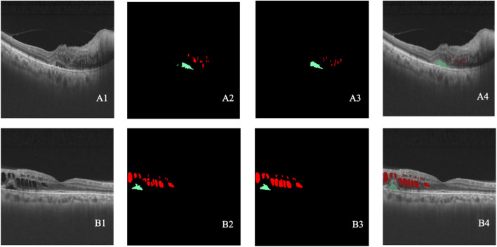
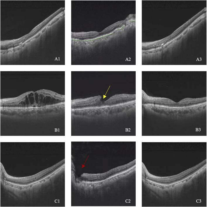
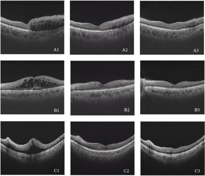
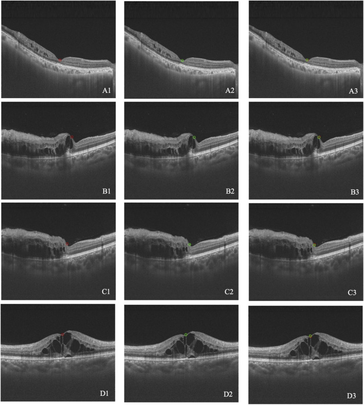
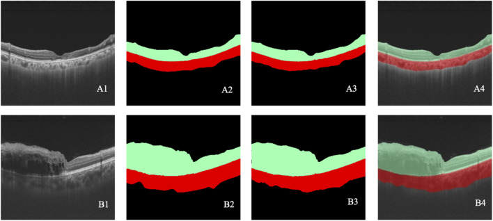
The study involved patients with RVO who received intravitreal anti-VEGF injection from 1 November 2018 to 30 November 2019. The OCT images taken before and shortly after treatment, with an interval of 4-8 weeks, were collected and randomly divided into the training set and test set at a ratio of approximately 3:1. The model is constructed based on the pix2pixHD algorithm, and synthetic OCT images are evaluated in terms of the picture quality, authenticity, the central retinal thickness (CRT), the maximal retinal thickness, the area of intraretinal cystoid fluid (IRC), and the area of subretinal fluid (SRF). Three supporting models, namely, the macular detection model, retinal stratification model, and lesion detection model, were constructed. Segmentation of macular location, retinal structure, and typical lesions were added to the input information. After verifying their accuracy, supporting models were used to detect the CRT, the maximal retinal thickness, IRC area, and SRF area of synthetic OCT images. The output predictive values are compared with real data according to the annotation on the real post-therapeutic OCT images.
A total of 1,140 pairs of pre- and post-therapeutic OCT images obtained from 95 RVO eyes were included in the study, and 374 images were annotated. Of the synthetic images, 88% were considered to be qualified. The accuracy of discrimination of real versus synthetic OCT images was 0.56 and 0.44 for two retinal specialists, respectively. The accuracy to predict the treatment efficacy of CRT, the maximal retinal thickness, IRC area, and SRF area was 0.70, 0.70, 0.92, and 0.78, respectively.
Our study proves that the GAN is a reliable tool to predict the therapeutic efficacy of anti-VEGF injections in RVO patients. Evaluations conducted both qualitatively and quantitatively indicated that our model can generate high-quality post-therapeutic OCT images. Consequently, it has great potential in predicting the treatment efficacy and providing guidance to clinical decision-making.
本研究旨在通过使用生成对抗网络(GAN),基于治疗前的光学相干断层扫描(OCT)图像生成治疗后的OCT图像。这些合成图像使我们能够预测视网膜静脉阻塞(RVO)患者玻璃体内注射抗血管内皮生长因子(VEGF)的短期治疗效果。
该研究纳入了2018年11月1日至2019年11月30日期间接受玻璃体内抗VEGF注射的RVO患者。收集治疗前和治疗后间隔4 - 8周拍摄的OCT图像,并按约3:1的比例随机分为训练集和测试集。基于pix2pixHD算法构建模型,并从图像质量、真实性、中心视网膜厚度(CRT)、最大视网膜厚度、视网膜内囊样液(IRC)面积和视网膜下液(SRF)面积等方面对合成的OCT图像进行评估。构建了三个辅助模型,即黄斑检测模型、视网膜分层模型和病变检测模型。将黄斑位置、视网膜结构和典型病变的分割添加到输入信息中。在验证其准确性后,使用辅助模型检测合成OCT图像的CRT、最大视网膜厚度、IRC面积和SRF面积。根据治疗后真实OCT图像上的标注,将输出预测值与真实数据进行比较。
本研究共纳入了从95只RVO眼睛获得的1140对治疗前和治疗后的OCT图像,其中374幅图像进行了标注。在合成图像中,88%被认为是合格的。两位视网膜专家对真实OCT图像与合成OCT图像的辨别准确率分别为0.56和0.44。预测CRT、最大视网膜厚度、IRC面积和SRF面积治疗效果的准确率分别为0.70、0.70、0.92和0.78。
我们的研究证明,GAN是预测RVO患者抗VEGF注射治疗效果的可靠工具。定性和定量评估均表明,我们的模型能够生成高质量的治疗后OCT图像。因此,它在预测治疗效果和为临床决策提供指导方面具有巨大潜力。