Bui Tim T, Yahanda Alexander T, Joseph Karan, Ruiz-Cardozo Miguel, de Monaco Bernardo A, Perdomo-Pantoja Alexander, Koleske Joshua P, McEvoy Sean D, Molina Camilo A
Department of Neurosurgery, Washington University School of Medicine, 660 S Euclid Ave, Campus Box 8057, St. Louis, MO 63110, USA.
Biomimetics (Basel). 2025 Jun 17;10(6):408. doi: 10.3390/biomimetics10060408.
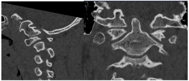
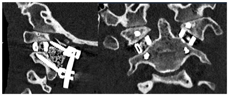
Type 1 basilar invagination (BI) is caused by a structural instability at the craniovertebral junction (CVJ) and has been historically treated with distraction and stabilization through fusion of the C1-C2 vertebrae. Recent advances in 3D printed custom implants (3DPIs) have improved the array of available options for reaching distraction and alignment goals. We report the case of a 15-year-old male who presented with early signs of cervical myelopathy. Radiographic evaluation revealed type 1 BI with a widened atlantodental interval (ADI) of 3.7 mm and a 9 mm McRae's line violation (MLV) of the dens, resulting in severe narrowing at the CVJ and brainstem/spinal cord impingement. Of note, the patient had bilateral dysplastic C1 and C2 anatomy, thus requiring a patient-specific 3DPI to conform to this anatomy and enable sufficient distraction and fusion. Custom 3D printed C1-C2 interfacet spacers were created and implemented within 14 days to achieve sufficient distraction, osteoconduction, and stabilization of the C1-C2 joint. Postoperatively, the patient remained neurologically intact with myelopathic symptom improvement before discharge on postoperative day 4. Postoperative imaging demonstrated the resolution of BI from successful C1-C2 joint distraction and confirmed intended implant placement with resolution of canal stenosis. During his 6-week follow-up, the patient remained neurologically stable with intact hardware and preserved alignment. This case is the first in the United States demonstrating the use of custom 3D printed interfacet spacers to achieve successful distraction, decompression, and stabilization of type 1 BI. These patient-specific 3DPIs were designed and created in a streamlined manner and serve as proof-of-concept of pragmatic implant design and manufacturing. Future optimization of the workflow and characterization of long-term patient outcomes should be explored for these types of 3DPI.
1型基底凹陷(BI)是由颅颈交界区(CVJ)的结构不稳定引起的,历史上一直通过C1-C2椎体融合进行牵引和稳定治疗。3D打印定制植入物(3DPI)的最新进展改善了实现牵引和对齐目标的可用选项。我们报告了一例15岁男性患者,其出现了颈椎病的早期症状。影像学评估显示为1型BI,寰齿间距(ADI)增宽至3.7mm,齿状突McRae线侵犯(MLV)达9mm,导致CVJ严重狭窄和脑干/脊髓受压。值得注意的是,该患者双侧C1和C2解剖结构发育异常,因此需要定制的3DPI以适应这种解剖结构并实现充分的牵引和融合。定制的3D打印C1-C2椎间关节间隔器在14天内制作并植入,以实现C1-C2关节的充分牵引、骨传导和稳定。术后,患者神经功能保持完整,脊髓病症状在术后第4天出院前有所改善。术后影像学显示BI通过成功的C1-C2关节牵引得到缓解,并确认植入物放置正确,椎管狭窄得到缓解。在6周的随访中,患者神经功能保持稳定,植入物完好,排列保持良好。该病例是美国首例展示使用定制3D打印椎间关节间隔器成功实现1型BI的牵引、减压和稳定的病例。这些定制的3DPI以简化的方式设计和制作,是实用植入物设计和制造的概念验证。对于这类3DPI,未来应探索工作流程的优化和长期患者预后的特征。