Eder Ann-Christin, Matthias Jessica, Lacombe Francois, Domogalla Lisa-Charlotte, Jacques Antoine, Steinacker Nils, Christien Gaetan, Martin Elodie, Criton Aline, Eder Matthias
Department of Nuclear Medicine, University Medical Center Freiburg, Faculty of Medicine, University of Freiburg, 79106 Freiburg, Germany.
Division of Radiopharmaceutical Development, German Cancer Consortium (DKTK), Partner Site Freiburg, Freiburg, Germany and German Cancer Research Center, 69120 Heidelberg, Germany.
Pharmaceuticals (Basel). 2025 Jun 4;18(6):841. doi: 10.3390/ph18060841.
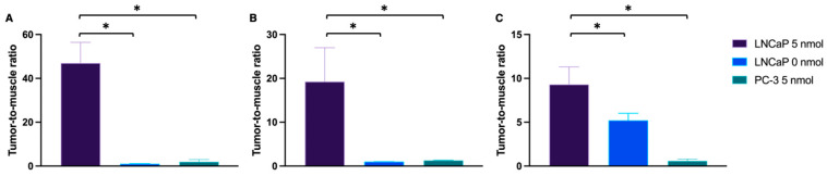
In prostate cancer (PCa) surgery, precise tumor margin identification remains challenging despite advances in surgical techniques. This study evaluates the combination of tumor-specific near-infrared imaging with the PSMA-targeting molecule PSMA-914 and optical endomicroscopy (NIR-pCLE) for single-cell-level tumor identification in a preclinical proof of concept. NIR-pCLE imaging of varying PSMA-914 concentrations was performed on PSMA-positive LNCaP and PSMA-negative PC-3 cells using Cellvizio 100 with pCLE Confocal Miniprobes™. To identify optimal PSMA-914 dosing for in vivo imaging, different doses (0-10 nmol) were evaluated using NIR-pCLE, Odyssey CLx imaging, and confocal microscopy in an LNCaP tumor-bearing xenograft model. A proof of concept mimicking a clinical workflow was performed using 5 nmol [Ga]Ga-PSMA-914 in LNCaP and PC-3 tumor xenografts, including PET/MRI, in/ex vivo NIR-pCLE imaging, and microscopic/macroscopic imaging. NIR-pCLE detected PSMA-specific fluorescence at concentrations above 30 nM in vitro. The optimal dose was identified as 5 nmol PSMA-914 for NIR-pCLE imaging with cellular resolution in LNCaP xenografts. PET/MRI confirmed high tumor uptake and a favorable distribution profile of PSMA-914. NIR-pCLE imaging enabled real-time, single-cell-level detection of PSMA-positive tissue, visualizing tumor heterogeneity, confirmed by ex vivo microscopy and imaging. This preclinical proof of concept demonstrates the potential of intraoperative PSMA-specific NIR-pCLE imaging to visualize tissue structures in real time at cellular resolution. Clinical implementation could provide surgeons with valuable additional information, potentially advancing PCa patient care through improved surgical precision.
在前列腺癌(PCa)手术中,尽管手术技术有所进步,但精确识别肿瘤边缘仍然具有挑战性。本研究在一项临床前概念验证中,评估了肿瘤特异性近红外成像与靶向PSMA的分子PSMA-914和光学内镜显微镜(NIR-pCLE)相结合用于单细胞水平肿瘤识别的情况。使用配备pCLE共聚焦微型探头™的Cellvizio 100对PSMA阳性的LNCaP细胞和PSMA阴性的PC-3细胞进行不同浓度PSMA-914的NIR-pCLE成像。为了确定体内成像的最佳PSMA-914剂量,在LNCaP荷瘤异种移植模型中使用NIR-pCLE、Odyssey CLx成像和共聚焦显微镜评估了不同剂量(0 - 10 nmol)。在LNCaP和PC-3肿瘤异种移植模型中,使用5 nmol [镓]镓-PSMA-914进行了模拟临床工作流程的概念验证,包括PET/MRI、体内/体外NIR-pCLE成像以及微观/宏观成像。NIR-pCLE在体外检测到浓度高于30 nM时的PSMA特异性荧光。对于LNCaP异种移植瘤的NIR-pCLE成像,确定最佳剂量为5 nmol PSMA-914以实现细胞分辨率。PET/MRI证实了PSMA-914的高肿瘤摄取和良好的分布情况。NIR-pCLE成像能够实时、单细胞水平检测PSMA阳性组织,可视化肿瘤异质性,这在体外显微镜检查和成像中得到了证实。这项临床前概念验证证明了术中PSMA特异性NIR-pCLE成像在细胞分辨率下实时可视化组织结构的潜力。临床应用可为外科医生提供有价值的额外信息,有可能通过提高手术精度推动PCa患者的治疗。