Wagner Nicole, Vukolic Ana, Baudouy Delphine, Pagnotta Sophie, Michiels Jean-Francois, Wagner Kay-Dietrich
CNRS, INSERM, iBV, Université Côte d'Azur, 06107 Nice, France.
Department of Cardiology, CHU Nice, 06107 Nice, France.
Theranostics. 2025 Jun 9;15(14):6593-6614. doi: 10.7150/thno.104329. eCollection 2025.
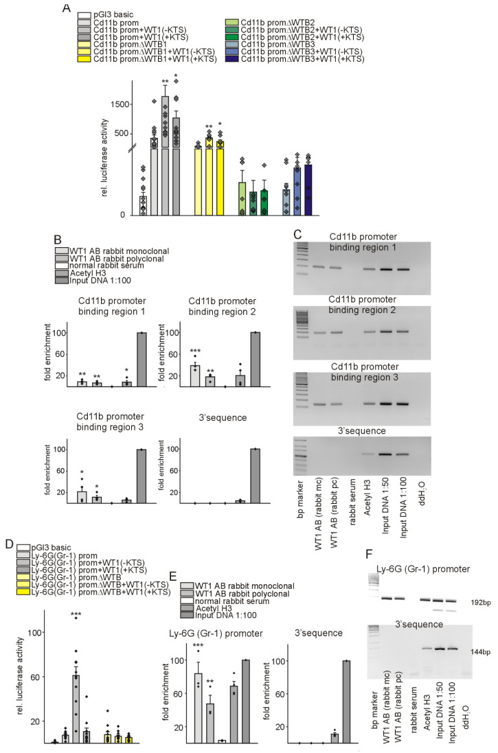
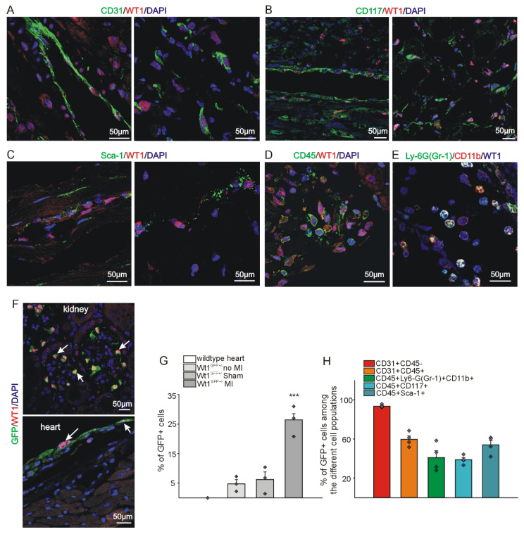
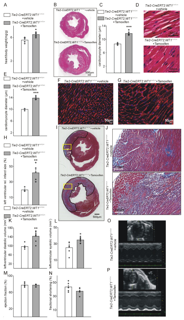
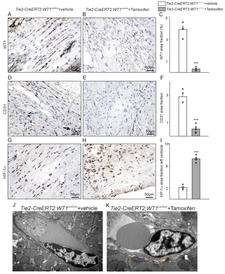
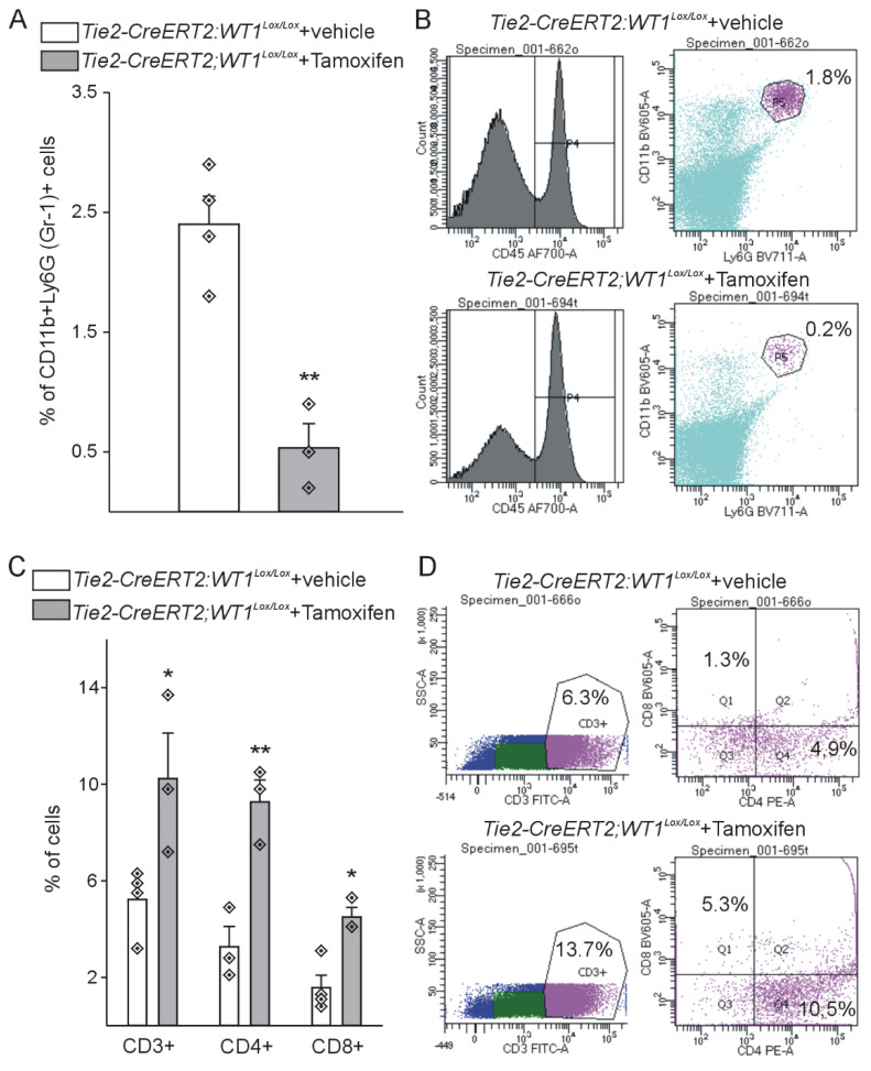
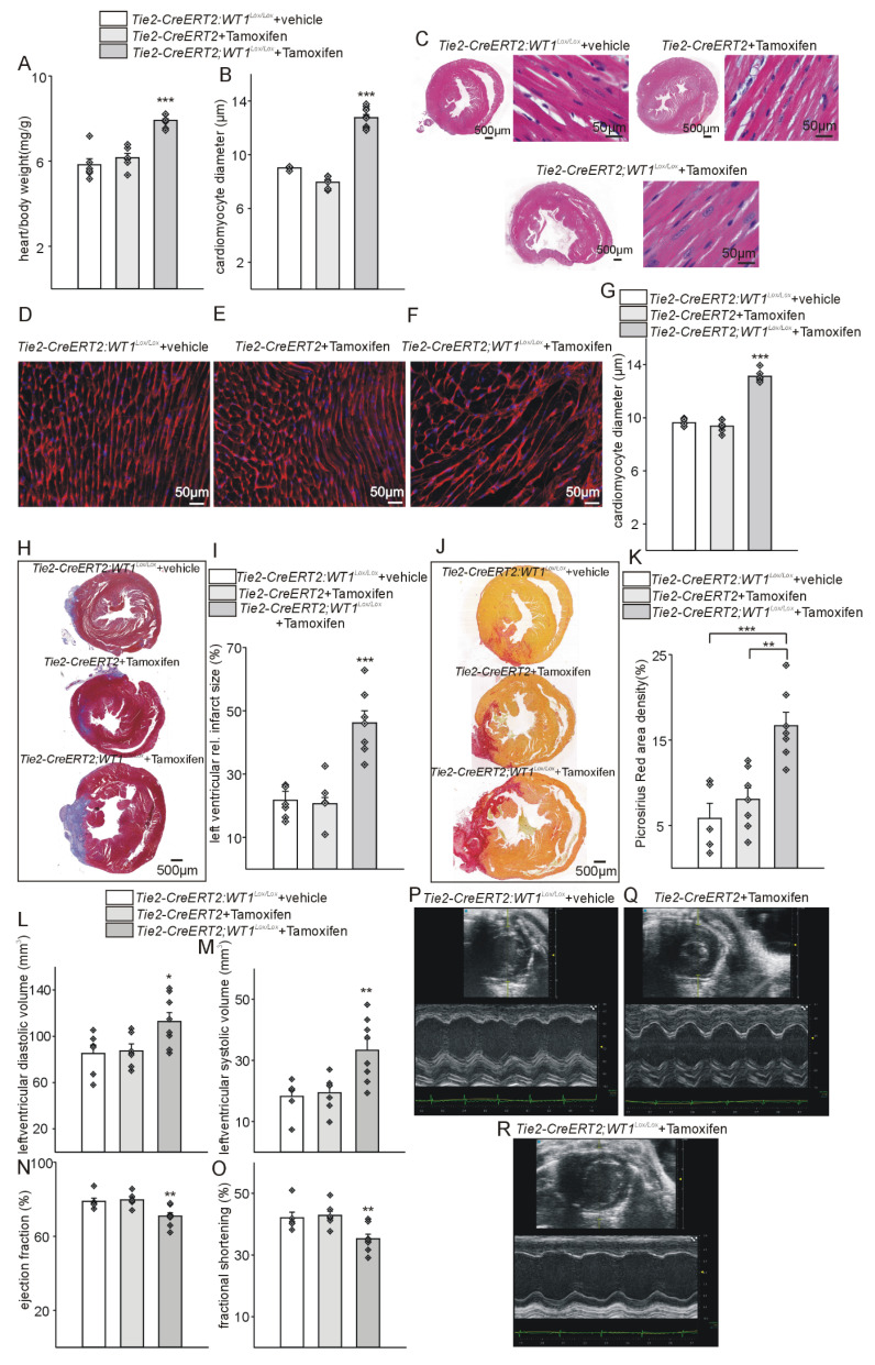
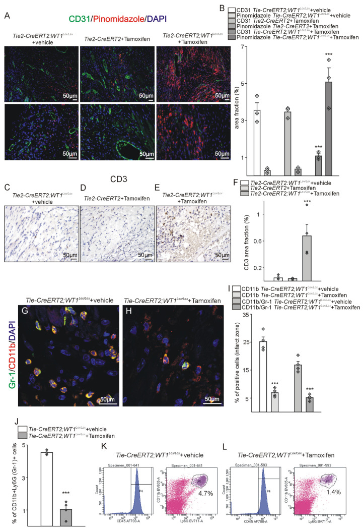
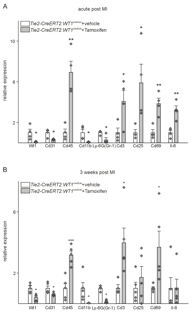
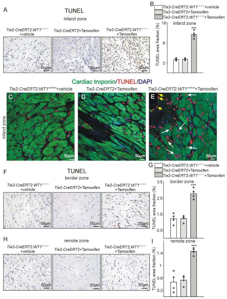
Cardiac repair and regeneration are severely constrained in adult mammals. Several cell types have been identified as playing a role in cardiac repair. However, our understanding of the regulatory proteins common to these cell types and implicated in cardiac repair remains limited. Experimental myocardial infarctions (MI) were induced in mice by ligation of the left coronary artery. WT1 expression in different cell types was determined by immunofluorescent double-labelling. VE-cadherin-CreERT2 (VE-CreERT2) mice were crossed with Wt1 animals to generate the VE-CreERT2;Wt1 strain to knockout WT1 in endothelial cells. Wt1 and Tie2-CreERT2 animals were crossed to generate Tie2-CreERT2;Wt1 mice to delete WT1 in endothelial and myeloid-derived cells. We show that the Wilms' tumor suppressor WT1 is expressed in progenitor cell populations, endothelial cells, and myeloid-derived suppressor cells (MDSCs) in mice following MI. Endothelial-specific knockout of WT1 results in reduced vascular density after MI but does not affect functional recovery. Conversely, combined knockout of WT1 in endothelial and myeloid-derived cells increases infarct size, cardiac hypertrophy, fibrosis, hypoxia, and lymphocyte infiltration. Notably, angiogenesis, infiltration of MDSCs, and cellular proliferation were diminished, and importantly, cardiac function was reduced. Mechanistically, in addition to the previously established role of WT1 in promoting the expression of angiogenic molecules, this transcription factor positively regulates the expression of Cd11b and Ly6G, which are crucial for MDSC invasion, migration and function thereby preventing overactivation of the immune response. Several molecules have been identified that are implicated in distinct aspects of cardiac repair following MI. The identification of WT1 as a transcription factor that is essential for repair mechanisms involving various cell types within the heart may potentially enable the future development of a coordinated repair process following myocardial infarction.
成年哺乳动物的心脏修复和再生受到严重限制。已确定几种细胞类型在心脏修复中发挥作用。然而,我们对这些细胞类型共有的、与心脏修复相关的调节蛋白的了解仍然有限。通过结扎左冠状动脉在小鼠中诱导实验性心肌梗死(MI)。通过免疫荧光双标记确定不同细胞类型中WT1的表达。将VE-钙黏蛋白-CreERT2(VE-CreERT2)小鼠与Wt1动物杂交,以产生VE-CreERT2;Wt1品系,从而在内皮细胞中敲除WT1。将Wt1和Tie2-CreERT2动物杂交,以产生Tie2-CreERT2;Wt1小鼠,从而在内皮细胞和髓系来源的细胞中删除WT1。我们发现,在MI后的小鼠中,肾母细胞瘤抑制因子WT1在祖细胞群体、内皮细胞和髓系来源的抑制细胞(MDSC)中表达。WT1的内皮特异性敲除导致MI后血管密度降低,但不影响功能恢复。相反,在内皮细胞和髓系来源的细胞中联合敲除WT1会增加梗死面积、心脏肥大、纤维化、缺氧和淋巴细胞浸润。值得注意的是,血管生成、MDSC浸润和细胞增殖减少,重要的是,心脏功能降低。从机制上讲,除了WT1在促进血管生成分子表达方面先前确立的作用外,这种转录因子还正向调节Cd11b和Ly6G的表达,这对于MDSC的侵袭、迁移和功能至关重要,从而防止免疫反应过度激活。已经确定了几种与MI后心脏修复的不同方面相关的分子。将WT1鉴定为一种转录因子,它对于涉及心脏内各种细胞类型的修复机制至关重要,这可能潜在地促进未来心肌梗死后协调修复过程的发展。