• 文献检索
  • 文档翻译
  • 深度研究
  • 学术资讯
  • Suppr Zotero 插件Zotero 插件
  • 邀请有礼
  • 套餐&价格
  • 历史记录
应用&插件
Suppr Zotero 插件Zotero 插件浏览器插件Mac 客户端Windows 客户端微信小程序
定价
高级版会员购买积分包购买API积分包
服务
文献检索文档翻译深度研究API 文档MCP 服务
关于我们
关于 Suppr公司介绍联系我们用户协议隐私条款
关注我们

Suppr 超能文献

核心技术专利:CN118964589B侵权必究
粤ICP备2023148730 号-1Suppr @ 2026

文献检索

告别复杂PubMed语法,用中文像聊天一样搜索,搜遍4000万医学文献。AI智能推荐,让科研检索更轻松。

立即免费搜索

文件翻译

保留排版,准确专业,支持PDF/Word/PPT等文件格式,支持 12+语言互译。

免费翻译文档

深度研究

AI帮你快速写综述,25分钟生成高质量综述,智能提取关键信息,辅助科研写作。

立即免费体验

内皮细胞中 Wt1 的缺失破坏了冠状动脉生成和心肌发育。

Endothelial deletion of Wt1 disrupts coronary angiogenesis and myocardium development.

机构信息

Celltec-UB, Department of Cell Biology, Physiology, and Immunology, Faculty of Biology, University of Barcelona, Barcelona 08028, Spain.

Institute of Biomedicine (IBUB), University of Barcelona, Barcelona 08028, Spain.

出版信息

Development. 2023 Mar 15;150(6). doi: 10.1242/dev.201147. Epub 2023 Mar 27.

DOI:10.1242/dev.201147
PMID:36852644
原文链接:https://pmc.ncbi.nlm.nih.gov/articles/PMC10112914/
Abstract

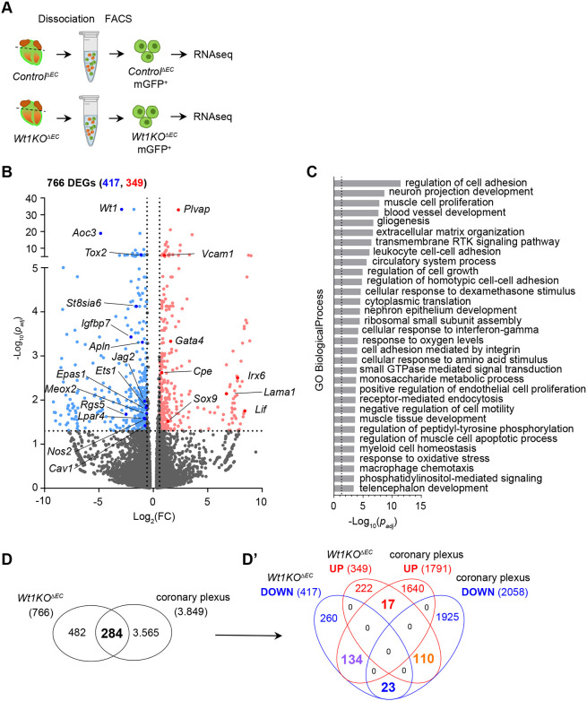
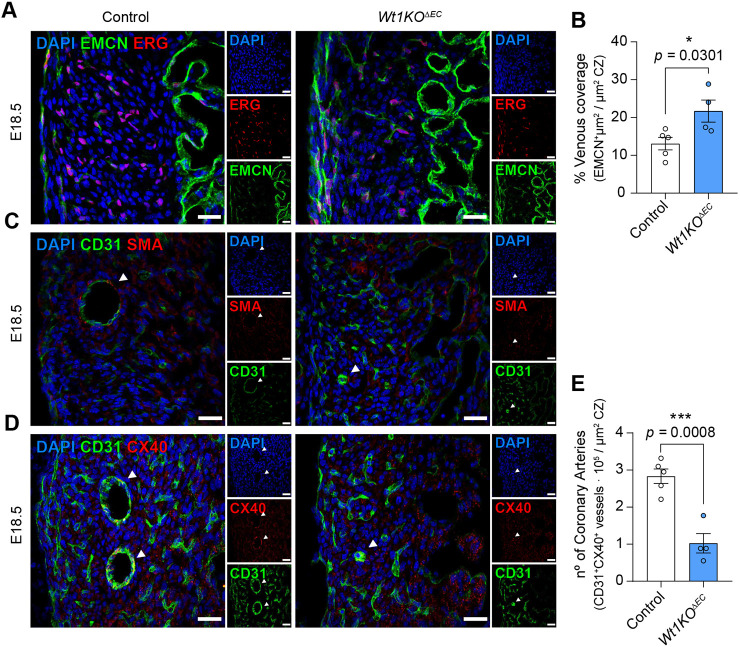
Wt1 encodes a zinc finger protein that is crucial for epicardium development. Although WT1 is also expressed in coronary endothelial cells (ECs), the abnormal heart development observed in Wt1 knockout mice is mainly attributed to its functions in the epicardium. Here, we have generated an inducible endothelial-specific Wt1 knockout mouse model (Wt1KOΔEC). Deletion of Wt1 in ECs during coronary plexus formation impaired coronary blood vessels and myocardium development. RNA-Seq analysis of coronary ECs from Wt1KOΔEC mice demonstrated that deletion of Wt1 exerted a major impact on the molecular signature of coronary ECs and modified the expression of several genes that are dynamically modulated over the course of coronary EC development. Many of these differentially expressed genes are involved in cell proliferation, migration and differentiation of coronary ECs; consequently, the aforementioned processes were affected in Wt1KOΔEC mice. The requirement of WT1 in coronary ECs goes beyond the initial formation of the coronary plexus, as its later deletion results in defects in coronary artery formation. Through the characterization of these Wt1KOΔEC mouse models, we show that the deletion of Wt1 in ECs disrupts physiological blood vessel formation.

摘要

WT1 编码一种锌指蛋白,对于心外膜的发育至关重要。尽管 WT1 也在心冠状内皮细胞(EC)中表达,但在 WT1 敲除小鼠中观察到的异常心脏发育主要归因于其在心外膜中的功能。在这里,我们生成了一种可诱导的内皮细胞特异性 WT1 敲除小鼠模型(Wt1KOΔEC)。在冠状动脉丛形成过程中内皮细胞中 WT1 的缺失损害了冠状动脉血管和心肌的发育。来自 Wt1KOΔEC 小鼠的冠状动脉 EC 的 RNA-Seq 分析表明,WT1 的缺失对冠状动脉 EC 的分子特征产生了重大影响,并改变了几个在冠状动脉 EC 发育过程中动态调节的基因的表达。这些差异表达的基因中的许多涉及冠状动脉 EC 的细胞增殖、迁移和分化;因此,上述过程在 Wt1KOΔEC 小鼠中受到影响。WT1 在冠状动脉 EC 中的作用超出了冠状动脉丛的初始形成,因为其后期缺失会导致冠状动脉形成缺陷。通过对这些 Wt1KOΔEC 小鼠模型的表征,我们表明 EC 中 WT1 的缺失破坏了生理性血管形成。

https://cdn.ncbi.nlm.nih.gov/pmc/blobs/3286/10112914/977e1995747e/develop-150-201147-g7.jpg
https://cdn.ncbi.nlm.nih.gov/pmc/blobs/3286/10112914/a1b7d56087e0/develop-150-201147-g1.jpg
https://cdn.ncbi.nlm.nih.gov/pmc/blobs/3286/10112914/c567978013a0/develop-150-201147-g2.jpg
https://cdn.ncbi.nlm.nih.gov/pmc/blobs/3286/10112914/39e22e8a6c8f/develop-150-201147-g3.jpg
https://cdn.ncbi.nlm.nih.gov/pmc/blobs/3286/10112914/d88ed9030454/develop-150-201147-g4.jpg
https://cdn.ncbi.nlm.nih.gov/pmc/blobs/3286/10112914/cd2653270563/develop-150-201147-g5.jpg
https://cdn.ncbi.nlm.nih.gov/pmc/blobs/3286/10112914/98b28d1c3d4a/develop-150-201147-g6.jpg
https://cdn.ncbi.nlm.nih.gov/pmc/blobs/3286/10112914/977e1995747e/develop-150-201147-g7.jpg

相似文献

1
Endothelial deletion of Wt1 disrupts coronary angiogenesis and myocardium development.内皮细胞中 Wt1 的缺失破坏了冠状动脉生成和心肌发育。
Development. 2023 Mar 15;150(6). doi: 10.1242/dev.201147. Epub 2023 Mar 27.
2
Cardiac endothelial cells express Wilms' tumor-1: Wt1 expression in the developing, adult and infarcted heart.心脏内皮细胞表达维尔姆斯瘤-1:Wt1 在心脏发育、成年和梗死中的表达。
J Mol Cell Cardiol. 2015 Apr;81:127-35. doi: 10.1016/j.yjmcc.2015.02.007. Epub 2015 Feb 11.
3
Coronary vessel development requires activation of the TrkB neurotrophin receptor by the Wilms' tumor transcription factor Wt1.冠状动脉发育需要威尔姆斯瘤转录因子Wt1激活TrkB神经营养因子受体。
Genes Dev. 2005 Nov 1;19(21):2631-42. doi: 10.1101/gad.346405.
4
The roadmap of WT1 protein expression in the human fetal heart.WT1蛋白在人胎儿心脏中的表达路线图。
J Mol Cell Cardiol. 2016 Jan;90:139-45. doi: 10.1016/j.yjmcc.2015.12.008. Epub 2015 Dec 10.
5
The Wilms' tumor suppressor Wt1 is expressed in the coronary vasculature after myocardial infarction.威尔姆斯瘤抑癌基因Wt1在心肌梗死后的冠状血管中表达。
FASEB J. 2002 Jul;16(9):1117-9. doi: 10.1096/fj.01-0986fje. Epub 2002 May 8.
6
The people behind the papers - Marina Ramiro-Pareta and Ofelia Martinez-Estrada.论文背后的人——玛丽娜·拉米罗 - 帕雷塔和奥费利娅·马丁内斯 - 埃斯特拉达。
Development. 2023 Mar 15;150(6). doi: 10.1242/dev.201740. Epub 2023 Mar 27.
7
Endocardial cushion morphogenesis and coronary vessel development require chicken ovalbumin upstream promoter-transcription factor II.心内膜垫形态发生和冠状血管发育需要鸡卵清蛋白上游启动子转录因子 II。
Arterioscler Thromb Vasc Biol. 2012 Nov;32(11):e135-46. doi: 10.1161/ATVBAHA.112.300255. Epub 2012 Sep 6.
8
Hippo Signaling Mediators Yap and Taz Are Required in the Epicardium for Coronary Vasculature Development.在冠状动脉血管发育过程中,心外膜需要Hippo信号通路的介质Yap和Taz。
Cell Rep. 2016 May 17;15(7):1384-1393. doi: 10.1016/j.celrep.2016.04.027. Epub 2016 May 5.
9
Experimental studies on the spatiotemporal expression of WT1 and RALDH2 in the embryonic avian heart: a model for the regulation of myocardial and valvuloseptal development by epicardially derived cells (EPDCs).WT1和RALDH2在鸡胚心脏中的时空表达实验研究:心外膜来源细胞(EPDCs)对心肌和房室间隔发育调控的模型
Dev Biol. 2002 Jul 15;247(2):307-26. doi: 10.1006/dbio.2002.0706.
10
Endothelial deletion of phospholipase D2 reduces hypoxic response and pathological angiogenesis.内皮细胞中磷脂酶 D2 的缺失可减少低氧反应和病理性血管生成。
Arterioscler Thromb Vasc Biol. 2014 Aug;34(8):1697-703. doi: 10.1161/ATVBAHA.114.303416. Epub 2014 Jun 19.

引用本文的文献

1
Cardiac Development, Cellular Composition and Function: From Regulatory Mechanisms to Applications.心脏发育、细胞组成与功能:从调控机制到应用
Cells. 2025 Sep 5;14(17):1390. doi: 10.3390/cells14171390.
2
Enhanced neoangiogenesis and balance of the immune response mediated by the Wilms' tumor suppressor WT1 favor repair after myocardial infarction.肾母细胞瘤抑癌基因WT1介导的新生血管生成增强及免疫反应平衡有利于心肌梗死后的修复。
Theranostics. 2025 Jun 9;15(14):6593-6614. doi: 10.7150/thno.104329. eCollection 2025.
3
Microenvironmental determinants of endothelial cell heterogeneity.

本文引用的文献

1
Perinatal angiogenesis from pre-existing coronary vessels via DLL4-NOTCH1 signalling.经 DLL4-NOTCH1 信号从预先存在的冠状动脉中进行围产期血管生成。
Nat Cell Biol. 2021 Sep;23(9):967-977. doi: 10.1038/s41556-021-00747-1. Epub 2021 Sep 8.
2
Coordination of endothelial cell positioning and fate specification by the epicardium.心外膜协调内皮细胞定位和命运特化。
Nat Commun. 2021 Jul 6;12(1):4155. doi: 10.1038/s41467-021-24414-z.
3
Arterialization requires the timely suppression of cell growth.动脉化需要及时抑制细胞生长。
内皮细胞异质性的微环境决定因素
Nat Rev Mol Cell Biol. 2025 Jun;26(6):476-495. doi: 10.1038/s41580-024-00825-w. Epub 2025 Jan 28.
4
The Wilms' Tumor Suppressor WT1 in Cardiomyocytes: Implications for Cardiac Homeostasis and Repair.心肌细胞中的肾母细胞瘤抑制因子WT1:对心脏稳态和修复的影响
Cells. 2024 Dec 17;13(24):2078. doi: 10.3390/cells13242078.
Nature. 2021 Jan;589(7842):437-441. doi: 10.1038/s41586-020-3018-x. Epub 2020 Dec 9.
4
Sox17 Controls Emergence and Remodeling of Nestin-Expressing Coronary Vessels.Sox17 控制巢蛋白表达的冠状血管的出现和重塑。
Circ Res. 2020 Nov 6;127(11):e252-e270. doi: 10.1161/CIRCRESAHA.120.317121. Epub 2020 Sep 14.
5
Coronary vessel formation in development and disease: mechanisms and insights for therapy.发育和疾病中的冠脉血管生成:治疗的机制与见解。
Nat Rev Cardiol. 2020 Dec;17(12):790-806. doi: 10.1038/s41569-020-0400-1. Epub 2020 Jun 25.
6
Dynamic Interstitial Cell Response during Myocardial Infarction Predicts Resilience to Rupture in Genetically Diverse Mice.心肌梗死后的动态间质细胞反应可预测遗传多样性小鼠对破裂的抵抗力。
Cell Rep. 2020 Mar 3;30(9):3149-3163.e6. doi: 10.1016/j.celrep.2020.02.008.
7
Coronary arterial development is regulated by a Dll4-Jag1-EphrinB2 signaling cascade.冠状动脉发育受 Dll4-Jag1-EphrinB2 信号级联调节。
Elife. 2019 Dec 4;8:e49977. doi: 10.7554/eLife.49977.
8
Epicardial cell shape and maturation are regulated by Wt1 via transcriptional control of .心外膜细胞的形状和成熟受 Wt1 调控,Wt1 通过. 的转录控制来实现。
Development. 2019 Oct 17;146(20):dev178723. doi: 10.1242/dev.178723.
9
Single-cell transcriptome analyses reveal novel targets modulating cardiac neovascularization by resident endothelial cells following myocardial infarction.单细胞转录组分析揭示了心肌梗死后驻留内皮细胞调节心脏新生血管的新靶点。
Eur Heart J. 2019 Aug 7;40(30):2507-2520. doi: 10.1093/eurheartj/ehz305.
10
The Wilms' tumor suppressor gene regulates pancreas homeostasis and repair.Wilms 瘤抑制基因调节胰腺稳态和修复。
PLoS Genet. 2019 Feb 14;15(2):e1007971. doi: 10.1371/journal.pgen.1007971. eCollection 2019 Feb.