Wang Qianru, Mo Xiumei, Li Hua, Ye Mingqiu, Fei Guojun, Chen Pinru, Wang Yongfei, Hou Xinpeng, He Jiajing, Liu Wenbin, Wang Jie, Yin Hui, Deng Zujun, Jin Xiaobao, Liu Zhenlong, Wang Qi, Huang Bo
Guangdong Provincial Key Laboratory of Pharmaceutical Bioactive Substances, Guangdong Pharmaceutical University, Guangzhou, 510006, People's Republic of China.
Department of Critical Care Medicine, Shenzhen Bao'an District Songgang People's Hospital, Shenzhen, 518105, China.
Parasit Vectors. 2025 Jul 1;18(1):249. doi: 10.1186/s13071-025-06863-3.
Cerebral malaria (CM), a fatal neurological complication of Plasmodium falciparum infection, is partially driven by neuronal injury. Emerging evidence highlights exosomes as vital mediators of mast cell-neuron interactions in neurological disease progression. While mast cells and their exosomes were previously shown to exacerbate experimental cerebral malaria (ECM) severity, the specific role of mast cell-derived exosomes in CM-associated neuronal injury remains unclear.
Exosomes were isolated from resting and lipopolysaccharide (LPS)-activated P815 mast cells (denoted as RE and AE, respectively) and characterized. These exosomes were administered to ECM mice and Plasmodium berghei ANKA (PbA)-infected red blood cell (iRBC)-stimulated neuronal HT-22 cells to investigate their functional impact and mechanisms.
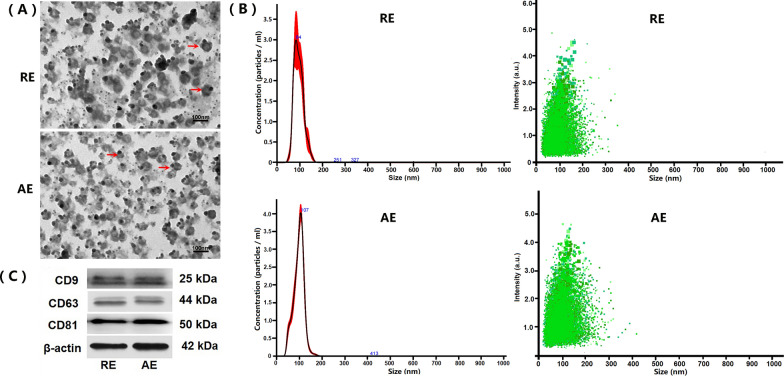
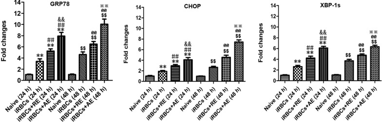
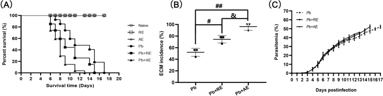
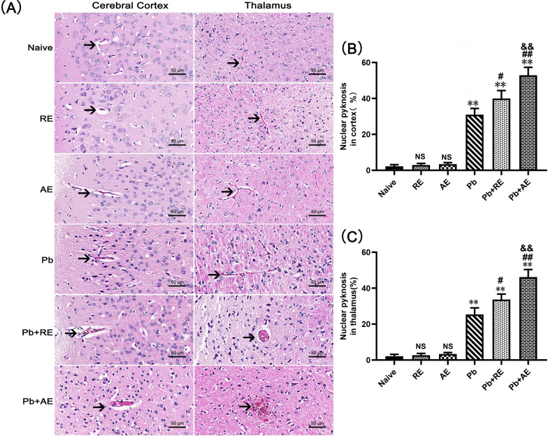
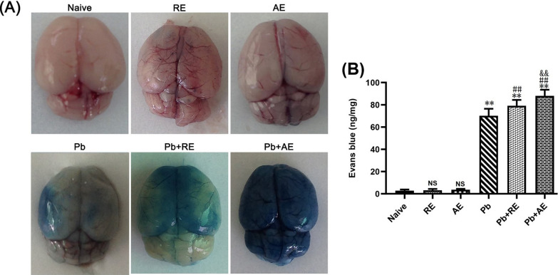
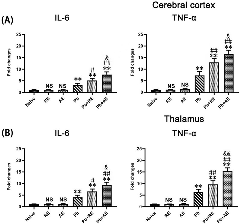
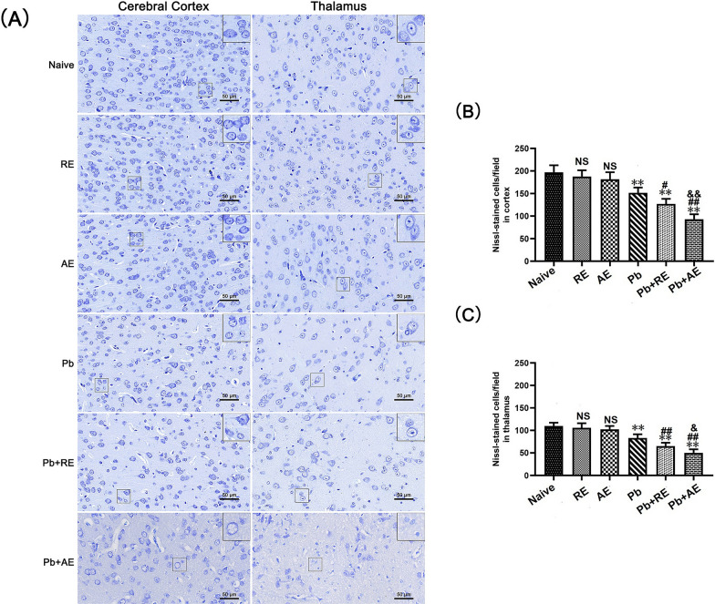
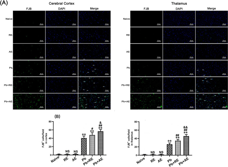
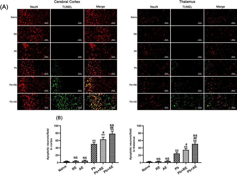
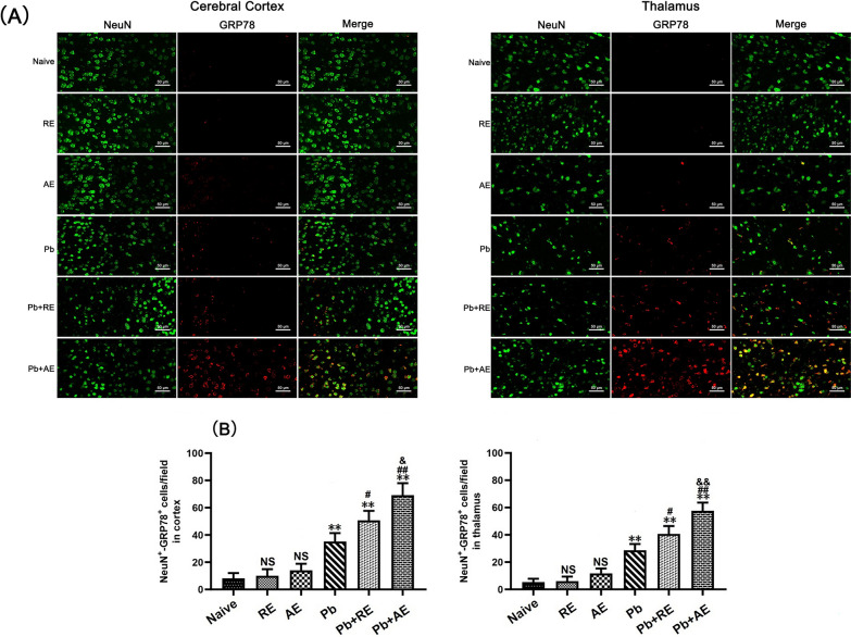
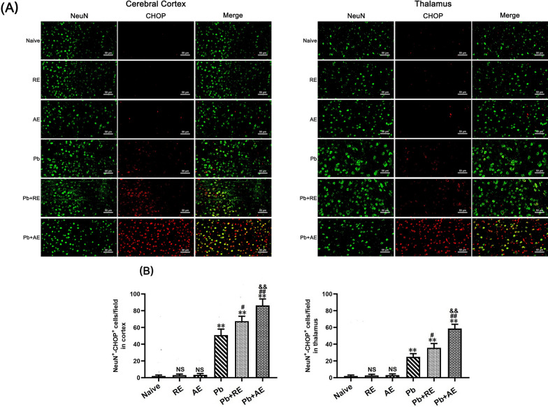
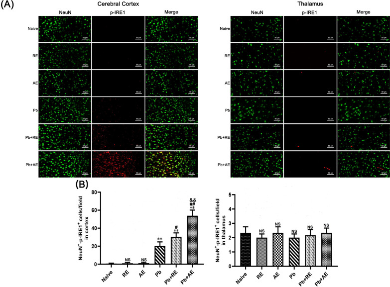
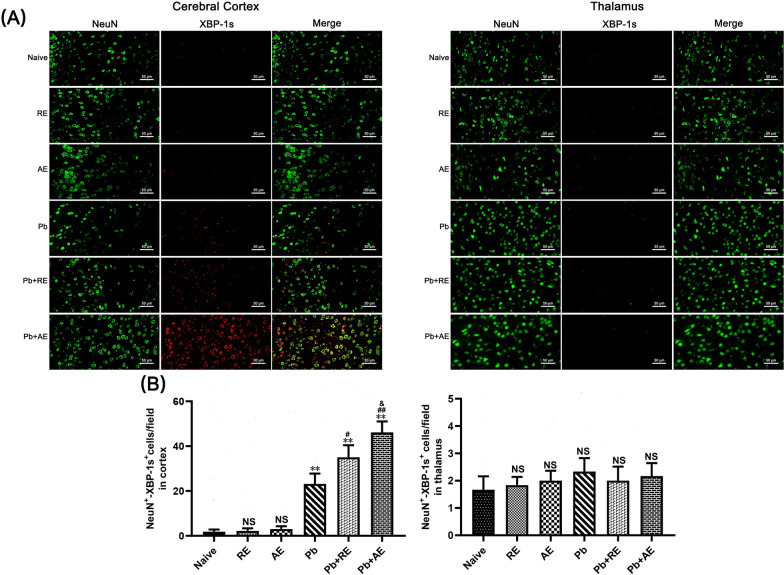
Both RE and AE exhibited spherical morphology (20-100 nm diameter) and expressed exosomal markers (CD9, CD63, and CD81). Compared to infected controls, RE and AE treatments significantly reduced survival time, increased ECM incidence, and exacerbated brain pathology, blood-brain barrier disruption, neuronal injury, and apoptosis. Furthermore, RE and AE administration elevated messenger RNA (mRNA) levels of pro-inflammatory cytokines (interleukin [IL]-6, tumor necrosis factor alpha [TNF-α], and IL-1β) and increased numbers of neurons expressing endoplasmic reticulum (ER) stress markers (GRP78, CHOP, p-IRE1, XBP-1). Notably, AE treatment induced higher morbidity/mortality rates, more severe neuronal injury, and greater ER stress marker expression than RE. In vitro, RE-treated iRBC-stimulated neuronal HT-22 cells showed higher GRP78, CHOP, and XBP-1 mRNA levels than AE-treated cells. MicroRNA (miRNA) sequencing revealed three downregulated miRNAs (miR-330-3p, miR-185-5p, and miR-379-5p) and six upregulated miRNAs (miR-155-5p, miR-423-3p, miR-187-3p, miR-29c-3p, miR-188-5p, miR-192-5p) in AE versus RE, all previously implicated in targeting GRP78, CHOP, or XBP-1.
Mast cell-derived exosomes, particularly those from activated cells (AE), exacerbated ECM neuronal injury through partial activation of ER stress pathways.
脑型疟疾(CM)是恶性疟原虫感染导致的一种致命性神经并发症,部分由神经元损伤所致。新出现的证据表明,外泌体是神经疾病进展过程中肥大细胞与神经元相互作用的重要介质。虽然肥大细胞及其外泌体先前已被证明会加重实验性脑型疟疾(ECM)的严重程度,但肥大细胞衍生的外泌体在与CM相关的神经元损伤中的具体作用仍不清楚。
从静息和脂多糖(LPS)激活的P815肥大细胞中分离外泌体(分别记为RE和AE)并进行表征。将这些外泌体给予ECM小鼠和感染伯氏疟原虫ANKA(PbA)的红细胞(iRBC)刺激的神经元HT-22细胞,以研究它们的功能影响和机制。
RE和AE均呈现球形形态(直径20 - 100 nm)并表达外泌体标志物(CD9、CD63和CD81)。与感染对照组相比,RE和AE处理显著缩短了生存时间,增加了ECM发病率,并加重了脑病理学改变、血脑屏障破坏、神经元损伤和细胞凋亡。此外,给予RE和AE提高了促炎细胞因子(白细胞介素[IL]-6、肿瘤坏死因子α[TNF-α]和IL-1β)的信使核糖核酸(mRNA)水平,并增加了表达内质网(ER)应激标志物(GRP78、CHOP、p-IRE1、XBP-1)的神经元数量。值得注意的是,AE处理比RE诱导了更高的发病率/死亡率、更严重的神经元损伤和更高的ER应激标志物表达。在体外,RE处理的iRBC刺激的神经元HT-22细胞显示出比AE处理的细胞更高的GRP78、CHOP和XBP-1 mRNA水平。微小核糖核酸(miRNA)测序显示,与RE相比,AE中有3种下调的miRNA(miR-330-3p、miR-185-5p和miR-379-5p)和6种上调的miRNA(miR-155-5p、miR-423-3p、miR-187-3p、miR-29c-3p、miR-188-5p、miR-192-5p),所有这些miRNA先前都与靶向GRP78、CHOP或XBP-1有关。
肥大细胞衍生的外泌体,尤其是来自活化细胞的外泌体(AE),通过部分激活ER应激途径加重了ECM神经元损伤。