Schürmann Till, Beschorner Ulrich, Westermann Dirk, Zeller Thomas, Bamberg Fabian, Schlett Christopher L, Stein Thomas, Noory Elias
Department of Diagnostic and Interventional Radiology, University Medical Center Freiburg, Faculty of Medicine, University of Freiburg, 79106, Freiburg, Germany.
Department of Cardiology and Angiology, University Heart Center, University Medical Center Freiburg, Faculty of Medicine, University of Freiburg, 79189, Bad Krozingen, Germany.
Sci Rep. 2025 Jul 9;15(1):24693. doi: 10.1038/s41598-025-06361-7.
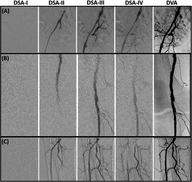
Digital variance angiography (DVA) exhibits promising prospects with respect to radiation exposure in digital subtraction angiography (DSA). This study aimed to determine the reduction of radiation dose in endovascular peripheral interventions (EPI) using DVA. The DVA imaging tool v6.0 (Kinepict Medical Imaging Tool, version 6.0.4, Kinepict Health Ltd., Budapest, Hungary) was utilized for patients undergoing EPI using digital angiography. EPI normal dose (EPI-ND) protocols were adapted from 1.20 to 0.81 µGy/frame to EPI low dose (EPI-LD) protocols using DVA-LD acquisitions with 0.36 µGy/frame and occasionally 0.24 µGy/frame based on specific examination requirements. The dose area product (DAP) was evaluated and contrast-to-noise ratio (CNR) was measured for each DSA acquisition. Evaluation included 370 EPI-ND and 62 EPI-LD using DVA-LD of three lower extremity regions (mean age: 73 ± 11 years, 67% male). LD protocols decreased median DAP of ND protocols significantly by 62.0% in pelvic, 53.8% in femoral and popliteal, and 59.4% in cruro-pedal regions, respectively (p < .005). DVA-LD increased median CNR significantly compared to DSA-LD (p < .001), and was equal to DSA-ND (p > .15). Image quality was enhanced by CNR/CNR ratio of 1.9 in pelvic, 2.4 in femoral and popliteal and in cruro-pedal regions. DVA reveals significant radiation dose reduction in lower extremity EPIs and enhances image contrast while decreasing noise.
数字方差血管造影术(DVA)在数字减影血管造影术(DSA)的辐射暴露方面展现出了广阔前景。本研究旨在确定使用DVA进行血管内周围介入治疗(EPI)时辐射剂量的降低情况。DVA成像工具v6.0(Kinepict医学成像工具,版本6.0.4,Kinepict健康有限公司,布达佩斯,匈牙利)被用于接受数字血管造影的EPI患者。EPI正常剂量(EPI-ND)方案从1.20微戈瑞/帧调整为EPI低剂量(EPI-LD)方案,使用DVA-LD采集,剂量为0.36微戈瑞/帧,根据特定检查要求偶尔为0.24微戈瑞/帧。对每次DSA采集评估剂量面积乘积(DAP)并测量对比噪声比(CNR)。评估包括三个下肢区域的370例EPI-ND和62例使用DVA-LD的EPI-LD(平均年龄:73±11岁,67%为男性)。LD方案使ND方案的中位DAP在盆腔区域显著降低62.0%,在股腘区域降低53.8%,在小腿足部区域降低59.4%(p<0.005)。与DSA-LD相比,DVA-LD显著提高了中位CNR(p<0.001),且与DSA-ND相当(p>0.15)。盆腔区域的CNR/CNR比值为1.9,股腘区域为2.4,小腿足部区域为2.4,图像质量得到增强。DVA显示下肢EPI的辐射剂量显著降低,在降低噪声的同时增强了图像对比度。