Balogh Boglárka, Zille Marietta, Szarka Gergely, Péntek Loretta, Futácsi Anett, Völgyi Béla, Kovács-Öller Tamás
Szentágothai Research Centre, University of Pécs, Pécs, Hungary.
Department of Neurobiology, Institute of Biology, Faculty of Sciences, University of Pécs, Pécs, Hungary.
Sci Rep. 2025 Jul 10;15(1):24804. doi: 10.1038/s41598-025-09007-w.
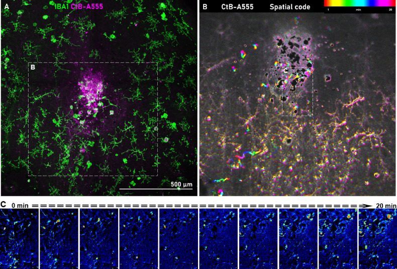
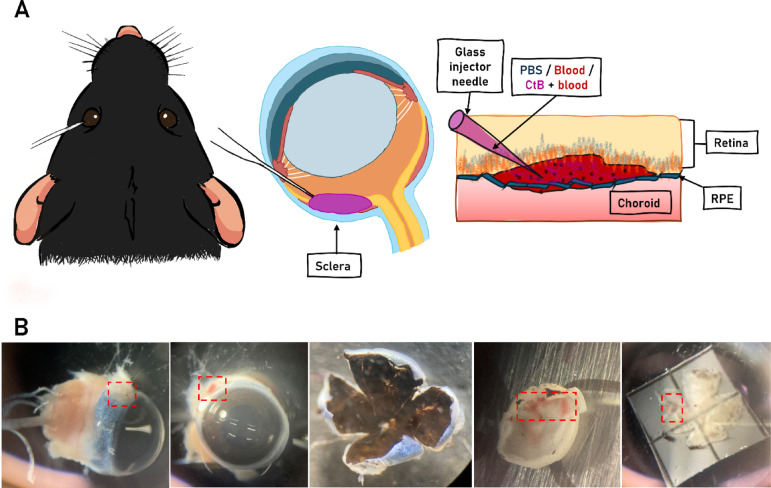
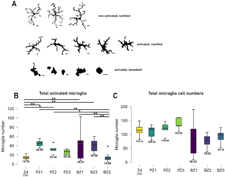
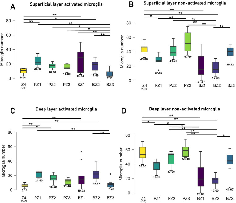
Subretinal hemorrhage (SRH) is caused by the accumulation of blood between the neurosensory retina and the retinal pigment epithelium or between the retinal pigment epithelium and the choroid. SRH often arises from age-related macular degeneration, traumas, and may occur spontaneously caused by other diseases like hypertension and diabetes. Here, we developed a novel technique - co-injection of blood and a dye-coupled tracer protein, Cholera toxin subunit B (CtB) - to better localize and understand the disease and how it can cause microglial activation, inflammation, and partial vision loss. Our results show that microglia are activated in the inner retinal layers in zones adjacent to blood injection. In contrast, the non-affected zone of the injected eye showed no microglial activation. For the first time, we used phosphate-buffered saline (PBS) injections as a control to assess the specific effects of injected blood. The results demonstrated that blood induced a markedly stronger activation response in the surrounding tissue, whereas PBS elicited a comparatively milder effect. PBS did cause microglial activation, but it was largely confined to the injection site and adjacent regions, and to a lesser extent than that observed with blood. We also observed microglial activation in the inner retina, along with the emergence of microglia and macrophages in the retinal pigment epithelium. Using advanced imaging techniques, we were able to better localize the affected area which comprises not only the immediate retinal area over the blood clot but the neighboring regions as well. These findings will provide the basis for novel therapeutic interventions targeting neuroinflammation in the retina after subretinal hemorrhage and other diseases affecting the eye.
视网膜下出血(SRH)是由血液在神经感觉视网膜与视网膜色素上皮之间或视网膜色素上皮与脉络膜之间积聚所致。SRH常源于年龄相关性黄斑变性、外伤,也可能由高血压和糖尿病等其他疾病自发引起。在此,我们开发了一种新技术——同时注射血液和一种染料偶联示踪蛋白霍乱毒素B亚基(CtB),以更好地定位和了解该疾病,以及它如何导致小胶质细胞活化、炎症和部分视力丧失。我们的结果表明,在与血液注射相邻区域的视网膜内层中,小胶质细胞被激活。相比之下,注射眼的未受影响区域未显示小胶质细胞活化。我们首次使用磷酸盐缓冲盐水(PBS)注射作为对照,以评估注射血液的特定作用。结果表明,血液在周围组织中诱导出明显更强的激活反应,而PBS引起的反应相对较轻。PBS确实会引起小胶质细胞活化,但主要局限于注射部位及相邻区域,且程度低于血液注射所观察到的情况。我们还观察到视网膜内层的小胶质细胞活化,以及视网膜色素上皮中出现小胶质细胞和巨噬细胞。使用先进的成像技术,我们能够更好地定位受影响区域,该区域不仅包括血凝块上方的直接视网膜区域,还包括邻近区域。这些发现将为针对视网膜下出血及其他影响眼睛的疾病后视网膜神经炎症的新型治疗干预提供依据。